|
HPI: An otherwise healthy 39-year-old female presents to the emergency department complaining of right shoulder pain. She had a fall the night previous where she fell directly onto the point of her right shoulder. She had dull, moderate pain but went to sleep after the fall as it was late and she had been drinking. She woke up on the morning of presentation with excruciating shoulder pain, inability to move her arm secondary to pain and a visible of the right shoulder deformity. No other traumatic injuries, no other complaints. She is not able to move her shoulder but can move her elbow and hand okay. No paresthesias, no weakness in the right arm. On physical exam, Her right shoulder is held against body, flexed at elbow. There is exquisite point tenderness over the AC joint. Appears deformed on visualization, with down-sloping of the anterior shoulder. Skin is intact. Palpation of the long bones and hands does not elicit tenderness or crepitus. The clavicle is neither tender nor deformed. Active/passive elbow and wrist range of motion is full and painless. Passive ROM of the right shoulder is not tolerated well secondary to pain. Patient can give a thumbs up, make an okay sign, and cross his index and long fingers without issue. Sensation to light touch is intact in the radial, median, and ulnar nerve distributions in the hand. Radial pulse is palpable. Right shoulder XRs were performed and are shown below. What is the diagnosis? Should you prepare the patient for shoulder reduction? Answer: AC Separation AC separation of the shoulder usually results from direct trauma to the AC joint, when the arm is in an adducted position. Support of the AC joint is through the acromioclavicular and coracoclavicular ligaments. Tenderness and deformity at the AC joint is diagnostic of this clinically. XRs are performed to confirm the diagnosis and also rule out underlying fracture. 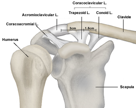 Image Credit: Orthobullets There are six types of AC separation. Involvement of the AC and CC ligaments determine the severity of acromio-clavicular injury. The types increasingly worsening in severity. 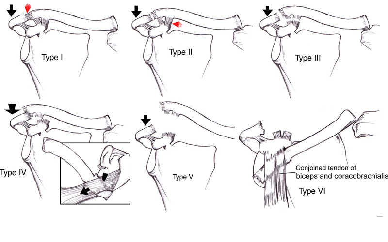 Image Credit: Orthobullets 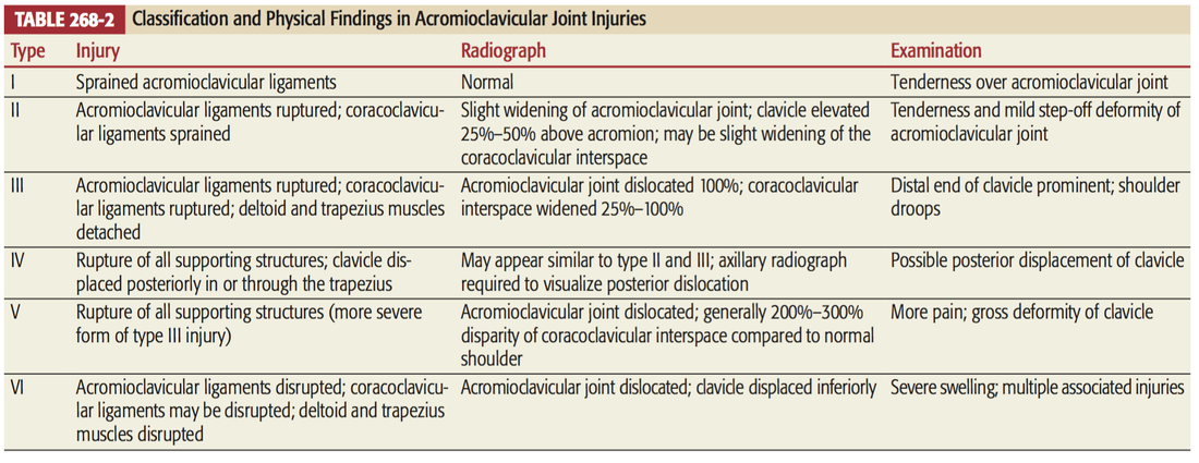 Image credit: Tintinalli's Radiographs: Must have True AP, Axillary and Scapular Y views to diagnose (to rule out associated dislocation or fracture). 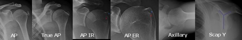 Image Credit: Orthobullets The normal AC joint space is 3mm and the normal coracoclavicular distance is 13 mm. Anything larger than these are pathologic. 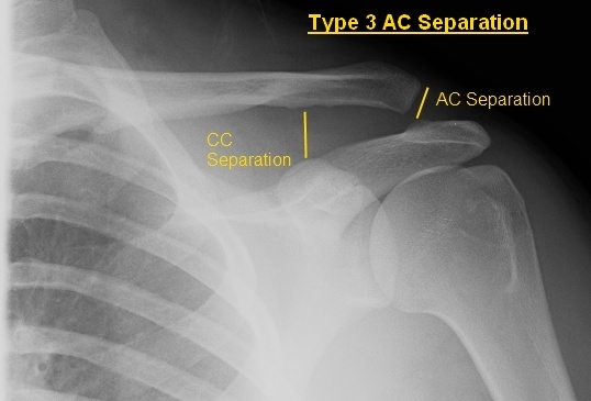 Image courtesy of: http://ortho-teaching.feinberg.northwestern.edu/cases/shoulder/case8/diagnosis.html%E2%80%8B Management:
Treatment of type I and II injuries consists of rest, ice, analgesics, and immobilization (sling), followed by early range-of-motion exercises (7 to 14 days). Orthopedic consultation for Types III through VI. Operative management is ORIF or ligament reconstruction: Type III in elite athletes, Types IV-VI. By: Dr. Michael Mollo M.D. References: 1. Rudzinski J Pittman L Uehara D: Injuries to Bones and Joints, in Tintinalli J., et al (eds): Tintinallis Emergency Medicine: A Comprehensive Study Guide, ed 7., (Sec) 22 (Ch) 268:p 1832-1834. 2. Orthobullets 3. Northwestern Orthopedics
0 Comments
HPI: Otherwise healthy middle-aged male presents to ED with L hand pain. He is a painter, and while cleaning a high-pressure pneumatic pain gun it accidentally discharged into the palm of his L hand. He reports only moderate pain at the site. Denies any other injuries, all vital signs within normal limits. Event occurred 2 hours prior to ED arrival. Exam: Pin-hole size puncture wound on palmar surface of hand w/ moderate tenderness to palpation and minimal surrounding erythema. Full passive/active ROM of all digits, neuromuscular function completely intact. Cap refill <2 seconds in all 5 digits. X-ray is unremarkable. 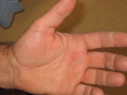 Discussion: High-Pressure Injection Injuries occur when a high-pressure injection device (pneumatic paint gun, grease gun, diesel injector, etc) injects into the operator. Most common in male laborers in painting and automotive industries, and injury often occurs during cleaning of the device or while attempting to clear the nozzle. Non-dominant hand involved ~75% of the time, with index finger injury most common, followed by middle finger and palm. Despite benign outward appearance, there is almost always significant underlying damage, and should be considered a SURGICAL EMERGENCY. Our job as ED providers is to recognize the seriousness of the injury and involve a hand surgeon as fast as possible! Pathophysiology involves direct trauma resulting in local tissue damage, acute and chronic inflammation, and foreign body granuloma formation. Tissue ischemia/necrosis from vascular compression, chemical inflammation, and secondary infection can lead to devastating functional outcomes, including amputation. Key historical details that impact prognosis:
Time from injury to treatment is the most important prognostic factor. Delay of >10 hours significantly increases risk of amputation. Higher PSI and larger volume of injected material portend greater direct tissue damage. Hydrocarbon-based substances (fuel, organic solvents, oil-based paint, paint thinners) cause more severe inflammatory reaction and tissue necrosis, with amputation rates >50%. Grease, latex, and water-based paints re typically less destructive. Obtain plain films in the ED to rule out coexistent fracture/dislocation. Some injected materials may appear radiopaque on x-ray and could aide in pre-operative planning. Administer broad-spectrum parenteral antibiotics in the ED, and update tetanus status. Control pain as needed, and arrange for emergent orthopedic/hand surgery evaluation. Transfer to Trauma Center if necessary. Patient will need emergent operative irrigation and debridement. Patient in HPI above had excellent functional outcome due to early presentation. Below is a time-course representation of patient with a delayed presentation, outlining potential damage that can occur. 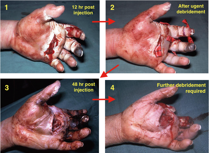 Management Pearls:
By Dr. Blake Johnson References: 1. Aiyer, A. "High-Pressure Injection Injuries." OrthoBullets, 15 Mar 2014. Web. Retrieved 25 Sept 2015, from http://www.orthobullets.com/hand/12104/high-pressure-injection-injuries. 2. Sanford, S. "High-Pressure Hand Injury." eMedicine/Medscape, 12 Nov 2013. Web. Retrieved 25 Sept 2015, from http://emedicine.medscape.com/article/826620-overview. (multiple images) 3. http://lifeinthefastlane.com/high-pressure-injection-injury/ (image) HPI: Otherwise healthy middle-aged male presents to ED with L wrist pain following moderate speed motorcycle collision. There was immediate pain/swelling to L wrist, and he is reluctant to range the joint. He was helmeted and wearing full protective clothing. Denies other injuries, all vital signs within normal limits. Exam: Prominent L wrist swelling. Dorsal aspect of radial head firmly palpated beneath the skin with slight volar displacement of hand relative to forearm. Skin intact, compartments are soft. Radial and ulnar pulses palpable, distal cap refill <2 seconds. Distal motor function intact, slightly diminished sensation in median nerve distribution of L hand. (X-ray shown below) 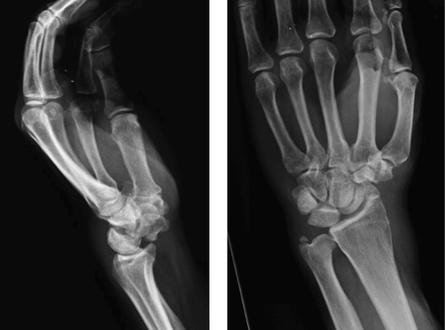 Radiologist Interpretation: Perilunate dislocation. Concomitant scaphoid waist fracture (central third) w/ distal anterior displacement. Minimally displaced ulnar styloid fracture. Discussion: Lunate dislocation (perilunate dissociation) is a high-energy injury with poor functional outcomes that almost universally requires operative intervention. Unfortunately they are commonly missed on initial presentation (~25%) due to subtleties on radiographic imaging. First, a review of normal carpal anatomy, characterized by 3 smooth lines called Gilula’s arcs: 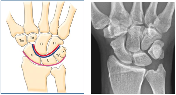 Any disruption in Gilula’s arcs raises suspicion for carpal fracture/dislocation. Lateral wrist film is essential to confirm normal in-line alignment of the radius, lunate, and capitate: 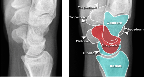 Mechanism of lunate dislocation typically involves high-energy axial load to the wrist, trapping the hand in hyperextension with ulnar deviation. Results in relative intercarpal supination causing varying degrees of carpal ligamentous rupture and articular dissociation. Injury pattern occurs in a step-wise sequence of events corresponding to Mayfield Classification System: · Stage I -> Scapho-lunate dissociation (SL widening) · Stage II -> Above w/ luno-capitate disruption (capitate overrides lunate) · Stage III -> Above w/ luno-triquetral disruption (true “perilunate dissociation”) · Stage IV -> Lunate completely dislocated from lunate fossa (usually volar, i.e. “spilled tea cup sign”) 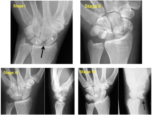 In addition to pain, stage IV dislocation and/or surrounding wrist fracture-dislocation can present with carpal tunnel compression and median nerve symptoms (~25% of patients). Emergent orthopedic consultation recommended. Closed reduction is performed in the ED using fingertraps to apply traction and to distract carpal bones while applying a sugar tong splint (may be all that is necessary). For complete lunate dislocations (stage IV), lunate must be relocated back into lunate fossa: 1) Apply gentle traction w/ wrist slightly extended 2) Manual pressure to palmar projection of lunate while flexing wrist until “snap” occurs (indicates relocation as proximal pole of capitate overcomes dorsal lip of the lunate) 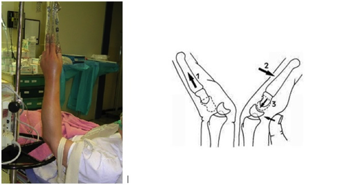 All acute lunate/perilunate dislocations require operative treatment with ORIF, ligament repair, and possible carpal tunnel release. There is universally poor functional outcomes with non-operative treatment, and recurrent dislocation is the rule, not the exception.
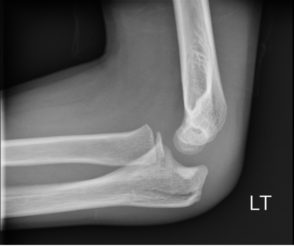
Management Pearls: · Know radiographic pathoanatomy, EASY TO MISS! · Assess for median nerve pathology and vascular injury · Emergent orthopedic consultation · Closed reduction with finger traps, apply sugar tong splint · Likely does not require admission, but operative treatment ASAP · High energy mechanism, don’t miss coexistent fracture or other injuries By Dr. Blake Johnson References: 1. Karadsheh, M. “Lunate Dislocation (Perilunate Dissociation).” OrthoBullets, 24 Dec 2014. Web. Retrieved 16 Sept 2015, from http://www.orthobullets.com/hand/6045/lunate-dislocation-perilunate-dissociation. (multiple images) 2. Murray, P. “Perilunate Fracture Dislocations.” eMedicine/Medscape, 22 Sept 2014. Web. Retrieved 16 Sept 2015, from http://emedicine.medscape.com/article/1240108-overview#a10. 3. http://radiologymasterclass.co.uk/tutorials/musculoskeletal/x-ray_trauma_upper_limb/wrist_trauma_x-ray.html (multiple images) 4. http://sfghed.ucsf.edu/Education/ClinicImages/Clin%20L%20finger%20trap%20w%20wts.1.jpg (image) 5. http://www.clicktocurecancer.info/kienbock-disease/ryan-j-grabow-mda-louis-catalano-iii-mdb.html (image) 6. Original case in HPI referred by Dr. Vivek Tayal HPI: Otherwise healthy middle-aged male presents to ED with L arm pain. He fell backwards off a ladder at work bracing the fall with his outstretched L hand. There was immediate pain/swelling to the elbow with inability to range the joint. He denies other injuries, and all vital signs are within normal limits. Exam: L arm adducted w/ elbow held in flexed position. Obvious swelling about elbow joint with prominent olecranon and shortened forearm. Compartments soft, distal motor/sensory function is intact. 2+ radial/ulnar pulses with cap refill <2 seconds. (x-ray shown below) 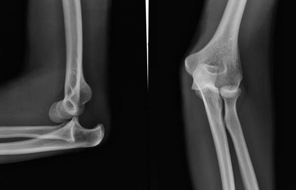 Discussion: Elbow dislocation is the second most common major joint dislocation seen in the ED (behind the shoulder). 80% of elbow dislocations are posterolateral and occur as a combination of: 1) axial loading, 2) forearm supination/external rotation, and 3) posterolateral valgus force to the elbow. · Simple dislocation - no associated fracture (approx 50-60% of cases). · Complex dislocation - one or multiple associated fractures present. Indications for Operative Repair include: 1) complex dislocation, 2) chronic dislocation, or 3) persistently unstable dislocation following attempted reduction. However, acute simple dislocation can typically be treated Non-Operatively w/ closed reduction and splinting. Multiple techniques for reduction exist: Reduction Technique #1 Prone position w/ elbow flexed at 90 degrees and humerus supported by the edge of the stretcher. Apply downward traction to patient’s forearm held in slight pronation, while using your other hand to apply downward and/or medial pressure to the olecranon. 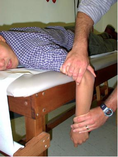 Reduction Technique #2 Supine or seated position w/ elbow held in slight flexion. Perform inline traction while supinating the forearm, with a second provider giving countertraction to the humerus if necessary. Gentle flexion of the elbow and/or medial pressure on the olecranon may be necessary to reduce lateral dislocations. 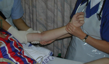 (YouTube video for demonstration: https://www.youtube.com/watch?v=mlAOGgocRnk)
After successful reduction, perform gentle ROM testing. Inability to range the joint smoothly raises suspicion for fracture, unsuccessful reduction, or re-dislocation. Full elbow extension is not necessary, as the joint is often still unstable in fully extended position. Place a posterior splint w/ forearm in slight pronation and elbow flexed at 90 degrees. Obtain post-reduction films, ensure soft compartments, and confirm intact neurovascular status. Orthopedic follow-up in 7-10 days. Obtain immediate orthopedic consultation for neurovascular compromise, fracture, or reduction failure. Management Pearls: · Plain films to diagnose dislocation and/or need for operative repair · Analgesia/anxiolysis (IV narcotics, hematoma block, +/- procedural sedation) · Successful reduction, gentle ROM, post-reduction films · Confirm intact neurovascular status · Posterior splint, immobilize 7-10 days, f/u with Orthopedics as outpatient By Dr. Blake Johnson References: 1. Aiyer A, Moore D. “Elbow Dislocation” [Web log post]. Retrieved 10 Sept 2015, from http://www.orthobullets.com/trauma/1018/elbow-dislocation. 2. Halstead, M. “Elbow Dislocation.” eMedicine/Medscape, 5 Aug 2014. Web. Retrieved 10 Sept 2015, from http://emedicine.medscape.com/article/96758-overview. 3. Case courtesy of Dr Maulik S Patel, Radiopaedia.org, rID: 14118. http://radiopaedia.org/articles/elbow-dislocation (image 1) 4. Video courtesy of Dr. Fakhouri, MidAmerica Orthopaedics and MidAmerica Hand To Shoulder Clinic. “Posterior Elbow Dislocation & Reduction.” https://www.youtube.com/watch?v=mlAOGgocRnk. 5. http://emedicine.medscape.com/article/96758-workup#c7 (image 2) 6. http://lifeinthefastlane.com/elbow-dislocation/ (image 3) HPI: Middle-aged male with no medical history presents to the ED with a laceration to his left middle fingertip. He was using a circular saw to cut wood this afternoon when he inadvertently cut his left middle finger. He denies other injuries, and all vital signs are within normal limits. Exam: Deep laceration to dorsal surface of distal phalanx on L middle finger involving the lateral nail bed. Full motor/sensory function is intact. Cap refill <2 seconds. Plain films negative for acute fracture/dislocation. (representative image seen below) 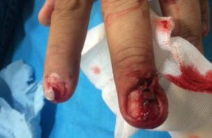 Discussion: Fingertip injuries are the most common hand injuries seen in the ED, and evidence shows effective early treatment clearly results in best cosmetic and functional outcomes. Special consideration is taken when injury involves the nail bed. · Germinal Matrix - soft tissue at base of the nail responsible for nail generation/growth. · Sterile Matrix - soft tissue adherent to underside of the nail plate responsible for nail strength/thickness. If nail plate is avulsed or subungual hematoma involves >50% of nail bed, then nail should be removed for hematoma evacuation/irrigation. If underlying laceration involves the germinal or sterile matrix, it should be repaired w/ 6-0 absorbable suture or Dermabond. Soak nail plate in a povidone iodine solution while performing repair. Replace the extracted nail plate at completion to splint the eponychial folds and better mold the edges of the repair for optimal healing (can be sutured in place). Apply a nonadherent dressing and consider a splint to immobilize DIP joint and protect the fingertip. 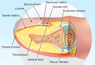 Management Pearls:
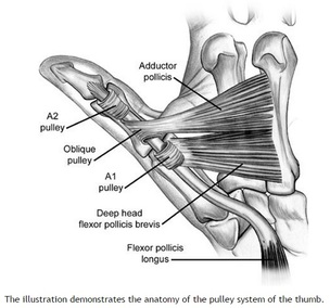
· Pain control (digital block), update tetanus status, Abx prophylaxis · Plain films to rule-out distal phalanx fracture · Copious irrigation and close inspection of wound · Nail plate removal and/or nail bed repair as indicated · Replace nail plate to splint eponychial fold and promote nail growth at germinal matrix · Non-urgent orthopedic follow-up as outpatient By Dr. Blake Johnson References: 1. Jones, T. Nail Bed Injury [Web log post]. Retrieved 3 Sept 2015, from http://www.orthobullets.com/hand/6109/nail-bed-injury. 2. Strauss EJ, Weil WM, Jordan C, et al. A prospective, randomized, controlled trial of 2-octylcyanoacrylate versus suture repair for nail bed injuries. J Hand Surg [Am]. 2008 Feb. 33(2):250-3. 3. Yallapragada, R. “Nail Bed Laceration Repair.” eMedicine/Medscape, 25 June 2015. Web. Retrieved 3 Sept 2015, from http://emedicine.medscape.com/article/80792-overview. 4. http://www.sawaccidents.com/table-saw-injury-pictures.htm (image 1) 5. http://www.medicinenet.com/image-collection/fingernail_anatomy_picture/picture.htm (image 2) HPI: Approximately 30 y/o right handed male presents with a laceration to base of the left thumb and inability to move his thumb. EXAM: Left hand: Laceration extends along the volar base of the thumb, no other injuries noted. Thumb is held in extension with limited opposition of the thumb. Complete inability to flex the thumb at the ITP. Ability to extend the thumb is preserved. When the wrist is extended, the thumb remains in extension. Decreased two point discrimination distal to the laceration. Ability to cross 2nd and 3rd digits is preserved. Remainder of sensation and motor function of the digits is intact. IMAGES: X-rays are needed only if underlying fracture is suspected (malalignment of digits)
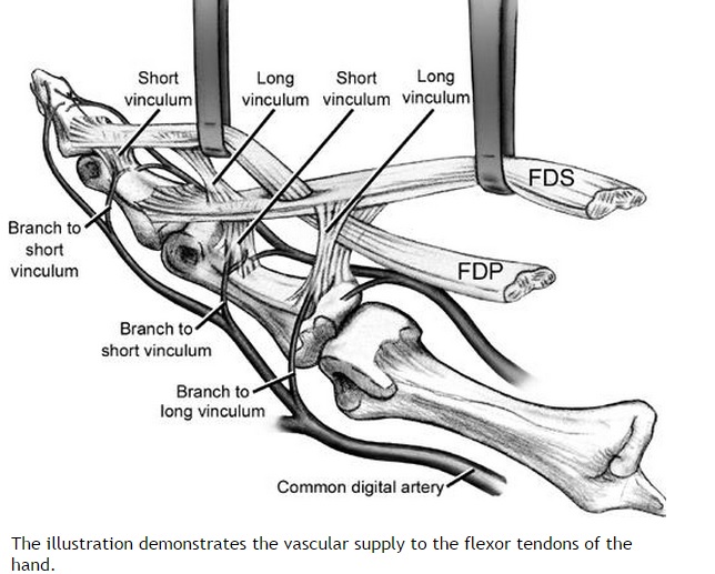
 MANAGEMENT
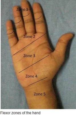
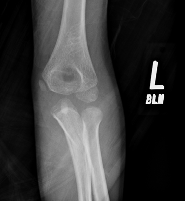
#ED Management - Assessment for other injuries - Thorough irrigation of wound and closure of skin - Splint in slight flexion - One dose of Ancef preferred - Very close follow up with hand specialist (1-3 days) #Non-Operative - <60% of tendon width affected - Wound care and hand rehab #Operative - >60% of tendon width affected - Ideally within 2 weeks of injury - Extensive hand rehab starting immediately after repair - Lots of repair techniques available depending on zone of injury. Outside the scope of this post DISCUSSION - This patient had a complete FPL transection. Laceration repaired, given 2 day f/u. - Flexor tendons need to be fixed ASAP but not emergently - More urgency than extensor tendon repairs - High suspicion with any volar laceration - Outcomes very variable depending on zone of injury and time until surgery - Hand rehabilitation is paramount to prevent scarring and loss of function - FDP inserts in the DIP. FDS inserts in the PIP. - High probability of concomitant neurovascular injury, associated with worse outcomes - Tenodesis effect - naturally, the digits flex when the wrist is extended. If abnormal very high likelihood of flexor tendon injury. KEY POINTS - High suspicion with volar lacerations and digits in extension - Remember the tenodesis effect - Always check for neurovascular injury - Higher urgency than extensor tendon injuries - Urgent but not emergent surgery required - Can close in the ED and arrange very close hand follow up - Splint in flexion - Be aware of the different different zones of the hand but extensive detail not needed. by Dr. Mohamed El-Kara REFERENCES: http://www.orthobullets.com/hand/6031/flexor-tendon-injuries HPI: 5 year old female presents with right arm pain after falling on a chair. Her arm was pinned in between the chair and the floor. She had immediate pain but it resolved. The following day she has increased pain and swelling in the right arm. EXAM: Swelling is noted in the proximal forearm. Tenderness to palpation present on the proximal ulna as well as radial head. No tenderness to the medial condyle, lateral condyle, or olecranon. Passive and active range of motion of the elbow is full but painful. No motor or sensory deficits of the hand. IMAGING. AP and lateral views of the elbow. 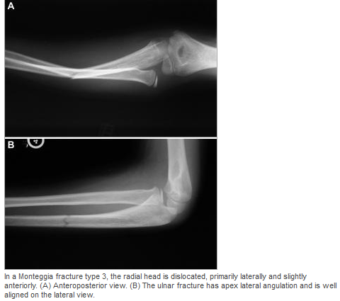 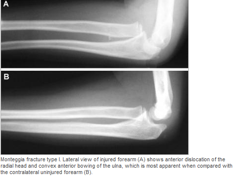 Note there is plastic deformation of the ulna without a complete fracture
Always assess the radial head, it should point towards the capitulum MANAGEMENT #Non-operative - Closed reduction of ulna and radial head dislocation and long arm casting - If no ortho available, attempt to reduce radial head and place in a posterior splint. - Axial traction traction to restore ulnar length - Need to successfully reduce radial head dislocation as well - Immobilize in 110 degrees flexion and full supination. #Operative - Indicated if radial head or ulnar length are unstable following reduction - Indicated in Bado Type IV fractures (complete fx of Ulna and Radius) - Frequently required in missed diagnosis. DISCUSSION - A Monteggia fracture is radial head dislocation plus a proximal ulna fracture or plastic deformation of the ulna - Peak age range 4-10 years, fall onto pronated arm.. - Frequently missed since the ulna may only have plastic deformation, have a high suspicion if there is pain over the radial head or evidence of radial head dislocation. - Complications include posterior interosseous nerve neurapraxia (finger drop, radial wrist deviation). - Loss of forearm motion with delayed treatment (2-3 weeks) - Different classifications are present (Bado) but not as key as recognizing the fracture. - This patient was casted and follow up arranged. KEY POINTS - Have a high index of suspicion when there is radial head tenderness or dislocation - Ulna may show only plastic deformation - Radial head should always point towards the capitulum - Obtain contralateral arm films if comparison is needed. - ED management without ortho includes reduction with posterior splint placement. Close f/u. - Majority of cases result in non-operative management. - Frequently missed By Dr. Mohamed El-Kara REFERENCES http://www.orthobullets.com/pediatrics/4015/monteggia-fracture--pediatric http://emedicine.medscape.com/article/415822-overview#a5 HPI: Right hand dominant middle aged female with history of HTN presents to the ED after accidentally hitting her left index finger with a hammer while working on a home improvement project. Finger is painful and swollen but she has no other injuries and reports she is otherwise feeling well. Physical Examination: Erythematous, swollen, and tender left 2nd distal phalanx. There is a subungual hematoma present over approx. 75% of the nail but the nail is intact. Motor and sensation intact, full ROM of PIP and DIP, 2+ radial pulse, no other injuries identified. Radiology: AP, lateral and oblique views of left 2nd phalanx – no fracture or dislocation identified Management: Nail removal and bedside repair of nail bed laceration, tetanus updated, discharged home with 48 hour follow up for reevaluation Discussion: Fingertip injuries = most common hand injuries seen in the ED Most common mechanisms:
Evaluate for:
Complications of injury:
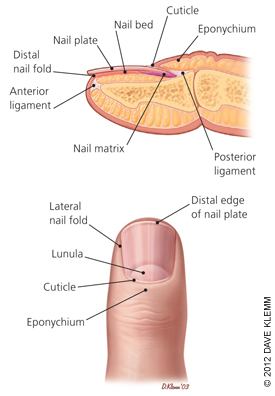 Image obtained from Am Fam Physician. 2012 Apr 15;85(8):779-787. Fingertip Anatomy:
Blood Supply and Innervation:
Nail Growth:
Treatment: Obtain radiographs to rule out distal phalanx fracture Evaluate for subunginal hematoma and nail bed lacerations Drain hematoma if <50% nail involved
Nail removal, I&D, repair of nail bed if >50% of nail involved
HPI: Young male otherwise healthy presenting today status post fall. Right hand dominant. Patient fell awkwardly with his arm slightly outstretched backwards off a trampoline and landed with his left arm under him. Patient had isolated pain at his left elbow. Ambulatory. No head injury or LOC. Physical Exam: Mild swelling present at the left elbow region with tenderness at the olecranon. Patient had limited range of motion at the elbow secondary to pain. Radial pulses strong and equal bilaterally. Able to give thumbs up, cross fingers, and make okay signs bilaterally. No neurologic deficits in median, radial or ulnar nerve distributions. Radiograph:
Management:
HPI:
Middle age male with no significant PMH presents to the ED after amputating his right index distal fingertip. Physical Examination: Patient has a right transverse (slightly volar oblique) index fingertip amputation with distal tuft visible within the wound. The laceration grazed the nailbed. Sensation is intact in the radial/medial/ulnar distributions of the hand including throughout the index finger. Radial pulse is palpable and the finger has good capillary refill. Radiology: XR of right hand is notable for distal tuft amputation but negative for foreign body. Management: Approximately 1mm of the nail plate was removed exposing the edge of the nailbed. Soft tissues, including the fat volarly and the nailbed dorsally were elevated off of the distal phalanx. Distal phalanx was then rongeured until sufficient soft tissue could be mobilized to cover the bone. The soft tissue was then sutured close. The wound was covered with Xeroform and dressed. Ancef was given prior to discharge. Orthopedic follow up was arranged for the following day. Discussion:
|
Orthopedics Blog
AuthorCMC ER Residents Archives
June 2018
Categories
All
Disclaimer: All images and x-rays included on this blog are the sole property of CMC EM Residency and cannot be used or reproduced without written permission. Patient identifiers have been redacted/changed or patient consent has been obtained. Information contained in this blog is the opinion of the author and application of material contained in this blog is at the discretion of the practitioner to verify for accuracy.
|
 RSS Feed
