CMC COMPENDIUM
  • Prospective Applicants
    • 2020 MATCH
    • About CMC
    • Our Curriculum
    • Our Residents
    • Our Fellowships
    • Program Leadership
    • Explore Charlotte
    • Official Site
  • ROTATING STUDENTS
    • Prospective Visiting Students
    • UNC Students
    • Healthcare Disparities Externship
    • Current Students
  • Current Residents
    • FlashPoint
    • Airway Lecture!
    • PGY - 1
    • PGY - 2
    • PGY - 3
    • Simulation Reading
    • Blogs >
      • EM GuideWire
      • CMC ECG Masters
      • Core Concepts
      • #FOAMed
      • Cardiology Blog
      • Dr. Patel's Coding Blog
      • Global Health Blog
      • Ortho Blog
      • Pediatric Emergency Medicine
      • Tox Blog
    • Resident Wellness
    • Resident Research
  • Top 20
  • Chiefs Corner
    • Schedules >
      • Conference/Flashpoint
      • Block Schedule
      • ED Shift Schedule
      • AEC Moonlighting
      • Journal Club/OBP/Audits Schedule
      • Simulation
    • Individualized Interactive Instruction
    • Evaluations/Interview Season
    • Contact Info
    • Resume Builder

Perilunate Dislocations

2/23/2015

0 Comments

 
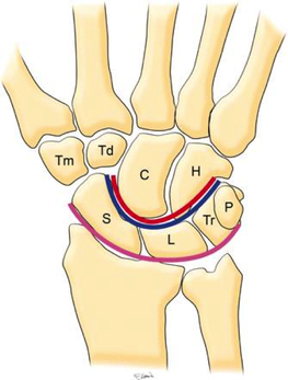
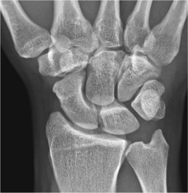
HPI: Young male presents after a fall while skiing with right shoulder and wrist pain.

PE: Obvious deformity of the right wrist. No abrasions or ecchymosis. Full thumb abduction, able to flex at DIP and PIP joint of all 5 fingers, full abduction of all 4 palmar fingers, 5/5 strength to finger grip. Unable to range wrist, elbow or glenohumeral joint secondary to pain. Normal sensation to pinprick and two point discrimination of all five fingers. 2+ radial pulse with normal cap refill.  

Imaging:
Picture
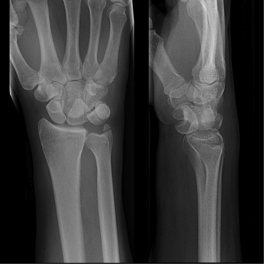
Anatomy:
-    Normal wrist anatomy consists of two rows of bones:
      o   Proximal row: Scaphoid, lunate, triquetrum, pisiform
      o   Distal row: trapezium, trapezoid, capitate, hamate
-    Ligaments of the wrist:
      o   Interosseous ligaments run between the carpal bones and stabilizes proximal carpal bones.
      o   Intrinsic ligaments insert and originate among the carpal bones to internally stabilize them.
      o   Extrinsic ligaments connect radius and ulna to the carpus. 
Picture
Picture
How to make diagnosis:
  • Traumatic high energy mechanism when wrist is extended and ulnarly deviated. 
  • Can lead to either Perilunate dislocation where lunate stays in position and carpus dislocates or Lunate dislocates by being forced volar or dorsal while carpus remains aligned
  • Can see median nerve symptoms in ~25% of patients – most common with lunate dislocation into carpal tunnel.

Treatment:
o   Require emergent closed reduction and splinting into sugar tong splint. 
  • Place hand in finger traps at 90 degrees of elbow flexion with 5-10lbs of traction. 
  • Wrist traction with one hand and recreate deformity (volar or dorsal dislocation) and pressing with thumb of the non-extending hand onto dislocated carpal bone attempting to press it back into place. 
  • Reduces median nerve injury and minimizes cartilage damage. 
o   Require open reduction, ligament repair, fixation and possible carpal tunnel release. 
  • Even with reduction and operative treatment may still not return to full function with reduced grip strength and stiffness. 

Pearls:
  • Must obtain a lateral to evaluate for fracture as AP may not reveal obvious carpal misalignment. 
  • Diagnosis missed 25% of the time. 
  • Either lunate stays in position and carpus dislocates or carpus stays in position and lunate dislocates. 
  • Emergent reduction/splinting to reduce incidence of median nerve injury and cartilage damage. 
0 Comments

Galeazzi Fracture

2/16/2015

0 Comments

 
HPI: Adult male presents after being involved in a motor vehicle collision with right wrist pain and obvious distal deformity.

PE: Obvious deformity of the right wrist. No abrasions or ecchymosis. Full thumb abduction, able to flex at DIP and PIP joint of all 5 fingers, full abduction of all 4 palmar fingers, 5/5 strength to finger grip. Slightly limited range of motion to flexion or extension wrist secondary to pain. Significant pain with pronation and supination. Normal sensation to pinprick and two point discrimination of all five fingers. 2+ radial pulse with normal cap refill. 

Imaging:
Picture
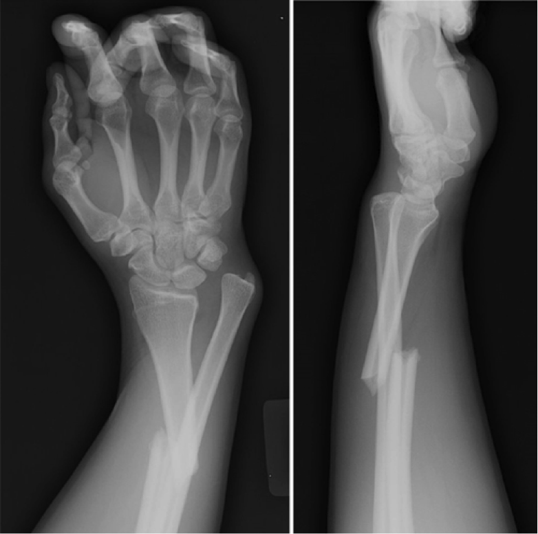
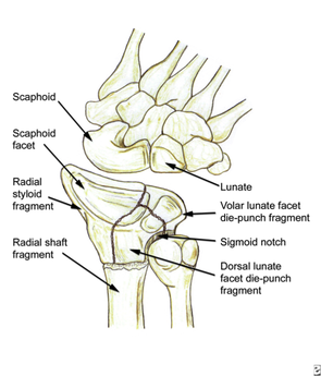
How to Diagnosis:

Radial shaft fracture and concurrent distal radioulnar joint. Most commonly seen in fractures of the distal 1/3 of the radius. Termed a Galeazzi fracture. Typically occurs secondary to direct wrist trauma (most classically dorsolateral) or with fall onto outstretched hand with forearm in pronation. 

Anatomy:
  • Radius and ulnar articulate along the sigmoid notch of the radius. 
  • Radioulnar ligaments along the volar and dorsal ligaments. 
  • This  relationship confers the stability of supination and pronation.
Picture
Treatment:
  • Requires ORIF of radius with reduction and stabilization of DRUJ. 

Pearls:
  • Key distance of radial fracture is 7.5cm from articular surface. DRUJ is unstable in 55% of fractures < 7.5cm, unstable in only 6% of fractures > 7.5cm. 
  • Concurrence of fracture and distal dislocation causes significant instability of supination and pronation.
  • Requires operative fixation, especially critical if fracture is < 7.5cm from articulating surface of radius. 

0 Comments

Ulnar Nerve Injury

2/8/2015

0 Comments

 
HPI:  Middle age male who presents after falling at work. His medial forearm stroke a metal bucket just distal to his elbow and resulted in a large laceration.

PE: Patient is unable to flex at the DIP of digit 4 and 5. He is unable to cross the second and third fingers or adduct his fingers. He has decreased sensation over the medial aspect of the 4th and 5th digit.  Suspected ulnar nerve laceration. 

Picture
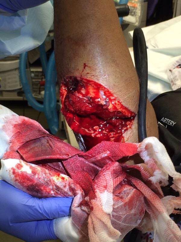
Anatomy: Derives from the medial portion of the brachial plexus (C8-T1). It lies posteromedial to the brachial artery in the upper arm and traverses behind the medial epicondyle.  The ulnar nerve runs along on the ulnar aspect of the wrist along with the ulnar artery. It passes through Guyon’s canal where it bifurcates into sensory and deep motor branches.  
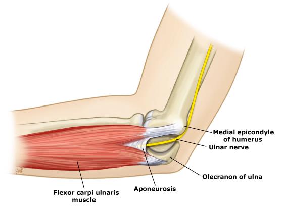
Picture
Innervation:
-   Motor:
  • Flexor carpi ulnaris and Flexor digitorum profundus for the 4th and 5th digits.
  • Adductor pollicis and deep head of flexor pollicis brevis
  • Interossei and 3rd and 4th lumbricals. 
  • Abductor digiti minimi, opponens digiti minimi, flexor digiti minimi. 

-    Sensory:
  •  Dorsal and palmar cutaneous branches as well as Superficial terminal branches. 

Clinical conditions:
  •  Distal humerus fracture
  •  External compression at the medial epicondyle
  •  Prolonged elbow flexion. 
  •  Medial epicondylitis (Golfer’s elbow)
  •  Compression in Guyon’s canal (spares 4th and 5th digit flexion)
  •  Propulsion of a wheelchair
  •  Fractures of the hook of the hamate

Treatment: Forearm exploration with transected nerve repair.

Pearls:
  •  Evaluate flexion at the DIP of the 4th and 5th digits. 
  •  Inablity to adduct the fingers. Crossing the 2nd and 3rd digit is maximum adduction. If unable to do this secondary to pain, can adduct 2nd and 3rd digit in “scissoring” motion against examiners fingers between the medial aspect of 2nd digit and lateral aspect of 3rd digit.  
  •  Decreased sensation at the lateral 4th and entire 5th digit. 
  •  Compression in Guyon’s canal will exhibit decreased sensation and inability to adduct the fingers. Flexion of 4th and 5th digit is spared.


0 Comments

    Orthopedics Blog

    Picture

    Author 

    CMC ER Residents 
    (for learning purposes, imagine all cases present to a free standing ED for dispo and treatment options)

    Subscribe To Blog

    Archives

    June 2018
    April 2018
    March 2018
    October 2017
    September 2017
    February 2016
    January 2016
    December 2015
    November 2015
    October 2015
    September 2015
    August 2015
    July 2015
    June 2015
    May 2015
    April 2015
    March 2015
    February 2015
    January 2015
    December 2014
    November 2014

    Categories

    All
    Lower Extremity
    Nerve Injury
    Spine
    Sports Medicine
    Upper Extremity
    Vascular Injury

    RSS Feed

    Disclaimer: All images and x-rays included on this blog are the sole property of CMC EM Residency and cannot be used or reproduced without written permission.  Patient identifiers have been redacted/changed or patient consent has been obtained.  Information contained in this blog is the opinion of the author and application of material contained in this blog is at the discretion of the practitioner to verify for accuracy.
Disclaimer: All original material and images included on this website are the sole property of CMC EM Residency and cannot be used or reproduced without written permission.  Information contained on this website is the opinion of the authors and does not necessarily represent the official opinion of Atrium Health or Carolinas Emergency Medicine Residency. 


For Health Care Providers:  Every effort is made to provide the most up to date evidence based medicine.  However, this content may not necessarily reflect the standard of care and application of material contained on this website is at the discretion of the practitioner to verify for accuracy.


For the Public:  This content is not intended to be a substitute for professional medical advice, diagnosis, or treatment.  Relying on information contained on this website is done at your own risk.  Do not disregard professional medical advice or delay seeking care secondary to content on this website.  Call 911 or seek a medical professional immediately for any medical emergencies.
Like us on Facebook or
follow us on Twitter/Instagram

Contact Us:

Department of Emergency Medicine
Medical Education Building., Third floor
1000 Blythe Blvd.
Charlotte, NC 28203

Telephone: 704-355-3658 
Fax: 704-355-7047
​
  • Prospective Applicants
    • 2020 MATCH
    • About CMC
    • Our Curriculum
    • Our Residents
    • Our Fellowships
    • Program Leadership
    • Explore Charlotte
    • Official Site
  • ROTATING STUDENTS
    • Prospective Visiting Students
    • UNC Students
    • Healthcare Disparities Externship
    • Current Students
  • Current Residents
    • FlashPoint
    • Airway Lecture!
    • PGY - 1
    • PGY - 2
    • PGY - 3
    • Simulation Reading
    • Blogs >
      • EM GuideWire
      • CMC ECG Masters
      • Core Concepts
      • #FOAMed
      • Cardiology Blog
      • Dr. Patel's Coding Blog
      • Global Health Blog
      • Ortho Blog
      • Pediatric Emergency Medicine
      • Tox Blog
    • Resident Wellness
    • Resident Research
  • Top 20
  • Chiefs Corner
    • Schedules >
      • Conference/Flashpoint
      • Block Schedule
      • ED Shift Schedule
      • AEC Moonlighting
      • Journal Club/OBP/Audits Schedule
      • Simulation
    • Individualized Interactive Instruction
    • Evaluations/Interview Season
    • Contact Info
    • Resume Builder