CMC COMPENDIUM
  • RESIDENCY
    • About CMC
    • Curriculum
    • Benefits
    • Explore Charlotte
    • Official Site
  • FELLOWSHIP
    • EMS
    • Global EM
    • Pediatric EM
    • Toxicology >
      • Tox Faculty
      • Tox Application
    • (All Others)
  • PEOPLE
    • Program Leadership
    • PGY-3
    • PGY-2
    • PGY-1
    • Alumni
  • STUDENTS/APPLICANTS
    • Medical Students at CMC
    • EM Acting Internship
    • Healthcare Disparities Externship
    • Resident Mentorship
  • #FOAMed
    • EM GuideWire
    • CMC Imaging Mastery
    • Pediatric EM Morsels
    • Blogs, etc. >
      • CMC ECG Masters
      • Core Concepts
      • Cardiology Blog
      • Dr. Patel's Coding Blog
      • Global Health Blog
      • Ortho Blog
      • Pediatric Emergency Medicine
      • Tox Blog
  • Chiefs Corner
    • Top 20
    • Current Chiefs
    • Schedules >
      • Conference/Flashpoint
      • Block Schedule
      • ED Shift Schedule
      • AEC Moonlighting
      • Journal Club/OBP/Audits Schedule
      • Simulation
    • Resources >
      • Fox Reference Library
      • FlashPoint
      • Airway Lecture
      • Student Resources
      • PGY - 1
      • PGY - 2
      • PGY - 3
      • Simulation Reading
      • Resident Wellness
      • Resident Research
      • Resume Builder
    • Individualized Interactive Instruction

Patella   fractures

6/28/2015

0 Comments

 
HPI: young male s/p ATV accident

Exam: Inspection: edema and deformity. No open wounds.  Palpation: TTP, palpable deformity. 2+ DP/PT pulses. SILT lower extremity. ROM: unable to perform straight leg raise. limited ROM secondary to pain. Limited assessment of anterior/posterior drawer, varus/valgus testing

Imaging:
 # Best seen on lateral xray. 
#In pediatric patients, MRI if xrays do not show fracture and child unable to perform straight leg raise
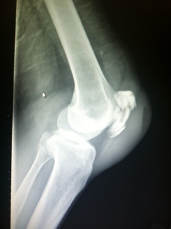
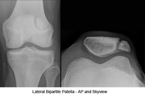
Picture
Pictureorthobullets.com


Bipartate patella: 
#often mistaken for fracture-use history and clinical exam
#8-10 % of population, 50 % bilateral, usually superolateral

Fracture Patterns:
  • nondisplaced
  • transverse
  • pole or sleeve (upper or lower)
  • vertical
  • marginal
  • osteochondral
  • comminuted (stellate)

Management:
# Nonoperative:
-Extensor mechanism intact (straight leg test)
-nondisplaced/minimally displaced
-vertical fractures
-Knee immobilized in extension (cylinder cast/brace)

#Operative
-extensor mechanism failure
-open fractures
-articular displacement > 2 mm
-displacement > 3 mm
-patella sleeve fractures in children (fracture between cartilage sleeve and patella)
-severely communited fractures

Discussion:
-Patella fractures 1 % of skeletal injuries
-Mechanism: direct impact of indirect eccentric contraction
-Complications: weakness and anterior knee pain, loss of reduction, nonunion, osteonecrosis, infection, stiffness

Key Points:  
#History of mechanism and exam is important-especially palpation, straight leg testing
#Best seen on lateral xray, consider MRI in pediatric patients given history and exam findings
#Bipartate patella-seen in 8-10 % of population, usually superolateral
#Orthopedic consultation-open vs closed, fracture pattern, straight leg testing, displacement
#Consider patella sleeve fractures in pediatric patients
#Management-minimally displaced, vertical, closed, extensor mechanism intact-immobilize in brace or cast and follow up as outpatient; Open, extensor mechanism not intact, communited, displaced-surgical

0 Comments

TOddler's  fractures

6/18/2015

0 Comments

 
HPI:
Toddler with history of osteogenesis imperfecta type 1 presented after a fall resulting in bruising to right lower extremity, fussiness. 

Exam:
vs normal
5 cm bruise to mid right shin, 2+ DP/PT pulses, cap refill < 2 sec. Full range of motion at hip, knee and ankle joint. Intact plantar, dorsiflexion, leg extension. NO laxity or pain with valgus or varus stress testing.

Imaging:
-Require AP and lateral views of tibia and fibula, ipsilateral knee and ankle
-Imaging below shows a non-displaced spiral tibial shaft fracture
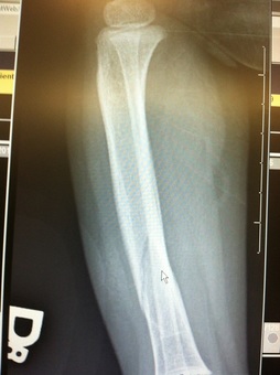
Picture
Management:
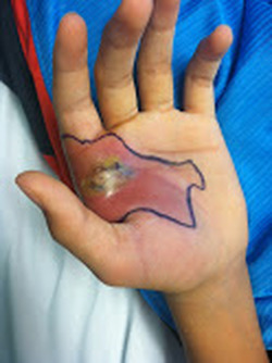
-Closed reduction with long leg cast
-Follow up with orthopedics in 2 weeks

Anatomy:
-Distal half of tibia, fibula usually remains intact

Discussion:
-15 % of all pediatric fractures
-If not walking yet, consider NAT
-May present with bruising, limping, refusal to bear weight
-Mechanism usually low energy with rotation

Key Points: 
-Toddler fractures common (15 %)
-Consider NAT if not walking yet
-AKA childhood accidental spiral tibia (CAST) fractures
-MOI: low energy, rotation
-History: pain, limp, not bearing weight
-Exam: warmth, pain, bruising, pain with ankle dorsiflexion
-Obtain: xrays AP/lateral tibia/fibula, ipsilateral knee and ankle
-Manage with closed reduction and long leg cast with orthopedic follow up in 2 weeks
-Prognosis: good, usually heal in 3-4 weeks
-Complications: compartment syndrome, leg-length discrepancy, angular deformity
0 Comments

Deep Space   Hand  infections

6/12/2015

0 Comments

 
HPI: Teenage  RHD healthy male presented initially to PCP with blister to left hand from playing baseball. Small blister incised and culture sent which eventually grew out MRSA and he was started on bactrim. On follow up 4 days later swelling worsened with worsening surrounding erythema. Denied fevers ,chills or other symptoms. Denied insect, spider or other exposures. Admitted for IV antibiotics and ortho hand consultation.

Exam: 
afebrile, vitals normal
2+ radial pulse, cap refill < 2 sec
SILT throughout hand and digits
Flexion/Extension/adduction/abduction intact and notable only for mild pain with flexion of thumb, area of fluctuance to radial aspect of palm extending from mid palm around to dosal aspect of 1st and 2nd digits; see picture
Picture
Picture
Imaging:
  • Plain films-if suspect foreign body
  • MRI-can provide information on extent of infection

Management:
Orthopedic hand consultation
Admission for IV antibiotics
  •  Unasyn-DOC
  • Ancef- alternative option
  • Vancomycin- if septicemia
  • Gentamicin- if penicillin allergic

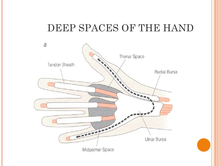
Anatomy:
3 main deep spaces: potential spaces separated by fascial septum
  • Thenar-bursa palmar to adductor pollicis and dorsal to flexor tendons
  • Deep Palmar-dorsal and radial to hypothenar space
  • Hypothenar-palmar from 5th metacarpal

Key Points:
  • Deep space infections include thenar (most common), hypothenar, midpalmar (rare)
  • Collar button abscess-between webspace of fingers
  • Exam reveals pain, swelling, pain with finger flexion
  • Differential Dx: paronychia, felon, flexor tenosynovitis
  • Imaging-xray for foreign body if suspected, consider MRI 
  • Consult orthopedic hand
  • Admission for IV antibiotics (unasyn DOC, alternatives include ancef, vancomycin, gentamicin


By: Dr Bryon Callahan
0 Comments

Yo, that subtalar joint looks gnarly! (subtalar  dislocations)

6/7/2015

0 Comments

 
Picture
Picture
Picture
HPI: 
Teenage male patient skating at high speed when his board abruptly came to a stop resulting in an inversion injury to his left ankle

PE: 
Skin intact


Left foot locked in supination with plantar flexion, obvious deformity to ankle

2+ DP/PT pulses, capillary refill < sec, SILT, 5/5 motor strength


Imaging: 
Medial subtalar dislocation: foot & calcaneus displaced medially, head of talus prominent dorsolaterally, navicular lies medial to talar head and neck


Lateral subtalar dislocation:  calcaneus displaced lateral to talus, talar head lies medially, navicular lies lateral to talar neck

Management:


·      Closed reduction-facilitated with knee flexion, relax gastrocnemius, may require sedation

·      Follow up CT scan ankle to assess for osteochondral lesions or fractures, assess reduction

·      Short leg cast/splint for 3-4 weeks

·      Approximately 10 % of medial dislocations and 20 % lateral require open reduction


Reduction Procedure:


https://www.youtube.com/watch?v=H6Dz566KrcM


 Anatomy: 
See attached

 Discussion:

·      Subtalar dislocation involves dislocation of distal articulations of talus at both talonavicular and talocalcaneal joints

·      Ankle joint undisturbed

·      Associated injuries: osteochondral lesions of talus, ankle fracture, fracture base of 5th metatarsal, navicular and cuboid fractures

·      Lateral dislocations less common, have more complications

Complications: infection (usually open), avascular necrosis (usually following fracture as talus is not disrupted from ankle mortoise and has blood supply).


     Key Points: 


·      Subtalar dislocations-medial more common than lateral, mechanism important part of HPI, clinical diagnosis

·      Obtain x rays to assess for associated injuries (ie fractures)

·      Manage with closed reduction and short leg cast/splint

·      10-20% may require open reduction

·      Obtain post reduction CT scan to assess reduction and osteochondral lesions/fractures

·      Complications rare but include infection (usually open) avascular necrosis ( usually associated fracture)

0 Comments

    Orthopedics Blog

    Picture

    Author 

    CMC ER Residents 
    (for learning purposes, imagine all cases present to a free standing ED for dispo and treatment options)

    Subscribe To Blog

    Archives

    June 2018
    April 2018
    March 2018
    October 2017
    September 2017
    February 2016
    January 2016
    December 2015
    November 2015
    October 2015
    September 2015
    August 2015
    July 2015
    June 2015
    May 2015
    April 2015
    March 2015
    February 2015
    January 2015
    December 2014
    November 2014

    Categories

    All
    Lower Extremity
    Nerve Injury
    Spine
    Sports Medicine
    Upper Extremity
    Vascular Injury

    RSS Feed

    Disclaimer: All images and x-rays included on this blog are the sole property of CMC EM Residency and cannot be used or reproduced without written permission.  Patient identifiers have been redacted/changed or patient consent has been obtained.  Information contained in this blog is the opinion of the author and application of material contained in this blog is at the discretion of the practitioner to verify for accuracy.
Powered by Create your own unique website with customizable templates.
  • RESIDENCY
    • About CMC
    • Curriculum
    • Benefits
    • Explore Charlotte
    • Official Site
  • FELLOWSHIP
    • EMS
    • Global EM
    • Pediatric EM
    • Toxicology >
      • Tox Faculty
      • Tox Application
    • (All Others)
  • PEOPLE
    • Program Leadership
    • PGY-3
    • PGY-2
    • PGY-1
    • Alumni
  • STUDENTS/APPLICANTS
    • Medical Students at CMC
    • EM Acting Internship
    • Healthcare Disparities Externship
    • Resident Mentorship
  • #FOAMed
    • EM GuideWire
    • CMC Imaging Mastery
    • Pediatric EM Morsels
    • Blogs, etc. >
      • CMC ECG Masters
      • Core Concepts
      • Cardiology Blog
      • Dr. Patel's Coding Blog
      • Global Health Blog
      • Ortho Blog
      • Pediatric Emergency Medicine
      • Tox Blog
  • Chiefs Corner
    • Top 20
    • Current Chiefs
    • Schedules >
      • Conference/Flashpoint
      • Block Schedule
      • ED Shift Schedule
      • AEC Moonlighting
      • Journal Club/OBP/Audits Schedule
      • Simulation
    • Resources >
      • Fox Reference Library
      • FlashPoint
      • Airway Lecture
      • Student Resources
      • PGY - 1
      • PGY - 2
      • PGY - 3
      • Simulation Reading
      • Resident Wellness
      • Resident Research
      • Resume Builder
    • Individualized Interactive Instruction