|
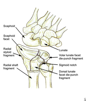
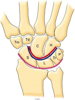
HPI: Young male presents after a fall while skiing with right shoulder and wrist pain. PE: Obvious deformity of the right wrist. No abrasions or ecchymosis. Full thumb abduction, able to flex at DIP and PIP joint of all 5 fingers, full abduction of all 4 palmar fingers, 5/5 strength to finger grip. Unable to range wrist, elbow or glenohumeral joint secondary to pain. Normal sensation to pinprick and two point discrimination of all five fingers. 2+ radial pulse with normal cap refill. Imaging: 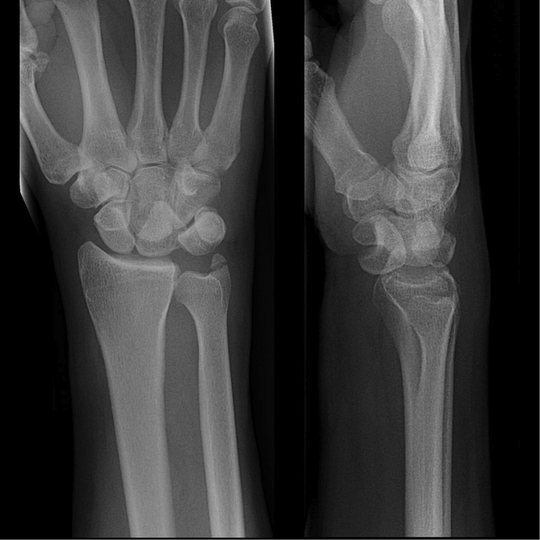 Anatomy: - Normal wrist anatomy consists of two rows of bones: o Proximal row: Scaphoid, lunate, triquetrum, pisiform o Distal row: trapezium, trapezoid, capitate, hamate - Ligaments of the wrist: o Interosseous ligaments run between the carpal bones and stabilizes proximal carpal bones. o Intrinsic ligaments insert and originate among the carpal bones to internally stabilize them. o Extrinsic ligaments connect radius and ulna to the carpus.
How to make diagnosis:
Treatment: o Require emergent closed reduction and splinting into sugar tong splint.
Pearls:
0 Comments
HPI: Adult male presents after being involved in a motor vehicle collision with right wrist pain and obvious distal deformity. PE: Obvious deformity of the right wrist. No abrasions or ecchymosis. Full thumb abduction, able to flex at DIP and PIP joint of all 5 fingers, full abduction of all 4 palmar fingers, 5/5 strength to finger grip. Slightly limited range of motion to flexion or extension wrist secondary to pain. Significant pain with pronation and supination. Normal sensation to pinprick and two point discrimination of all five fingers. 2+ radial pulse with normal cap refill. Imaging: 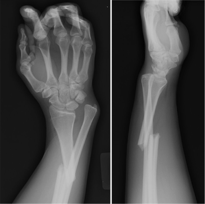
How to Diagnosis: Radial shaft fracture and concurrent distal radioulnar joint. Most commonly seen in fractures of the distal 1/3 of the radius. Termed a Galeazzi fracture. Typically occurs secondary to direct wrist trauma (most classically dorsolateral) or with fall onto outstretched hand with forearm in pronation. Anatomy:
Treatment:
Pearls:
HPI: Middle age male who presents after falling at work. His medial forearm stroke a metal bucket just distal to his elbow and resulted in a large laceration. PE: Patient is unable to flex at the DIP of digit 4 and 5. He is unable to cross the second and third fingers or adduct his fingers. He has decreased sensation over the medial aspect of the 4th and 5th digit. Suspected ulnar nerve laceration. 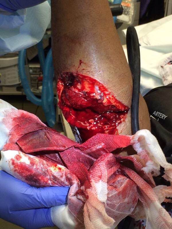 Anatomy: Derives from the medial portion of the brachial plexus (C8-T1). It lies posteromedial to the brachial artery in the upper arm and traverses behind the medial epicondyle. The ulnar nerve runs along on the ulnar aspect of the wrist along with the ulnar artery. It passes through Guyon’s canal where it bifurcates into sensory and deep motor branches. 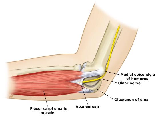 Innervation:
- Motor:
- Sensory:
Clinical conditions:
Treatment: Forearm exploration with transected nerve repair. Pearls:
HPI: Pediatric male presents after a fall onto outstretched arm with obvious deformity to right elbow. PE: Patient is unable to cross fingers and has paresthesias in the ulnar nerve distribution. Initial radiograph showed posterior dislocation with questionable hyperdensity representing the medial epicondyle. We were unable to reduce under fluoroscopy as the medial epicondyle was more visible and the patient eventually required ORIF. Imaging: AP and lateral radiograph of the elbow 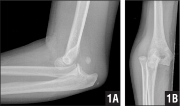
Image 1: Showing associated medial epicondyle fracture How to Diagnosis: Two important lines help in making the diagnosis; radiocapitella and anterior humeral. Radiocapitellar line: Drawn through the center of the radial neck and should pass the center of the capitellum as shown below 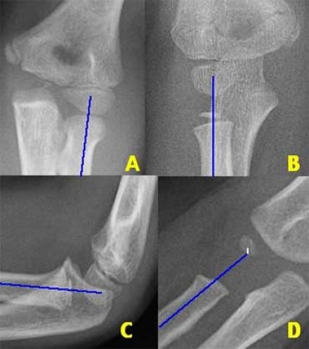
Anterior humeral line: Drawn through the lateral view of the surface of the humerus and should pass through the middle of the capitellum 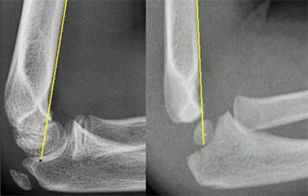
Ossification: 6 sites that fuse at different ages. Mnemonic = CRITOE
Treatment: Nonoperative with closed reduction and early range of motion (1-2 weeks) is most common. ORIF required for incarcerated medial epicondyle, inability to obtain close reduction, or significant instability. Pearls:
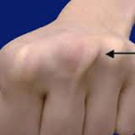
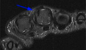
HPI: Young male presents after feeling a pop in his finger after he dropped a case of bottled water causing his middle finger to get caught in the wrapping. PE: Ulnar deviation of affected digit and a popping sensation and pain when flexing his fingers.
Imaging:
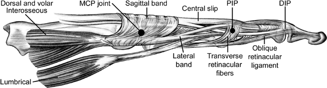
Anatomy:
Treatment Options:
HPI: 7 y/o fall from monkey bars. Landed on extended shoulder + outstretched arm. Physical exam: Obvious arm deformity. Ecchymosis over distal/medial arm. Inability to flex thumb IP joint and DIP of index finger (AIN neuropraxia). Palpable pulses. Warm extremity. 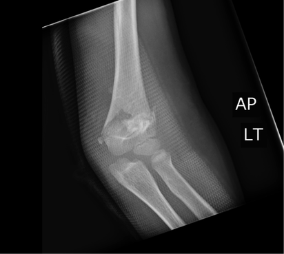
AP showing mild varus angulation 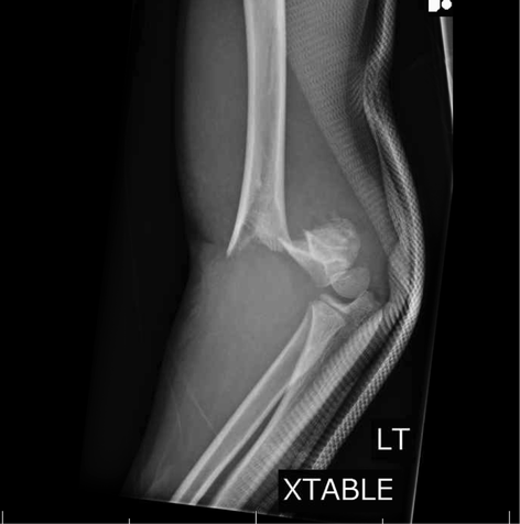
Lateral film showing significant posterior displacement of distal portion of fracture 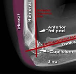
Normal lateral film 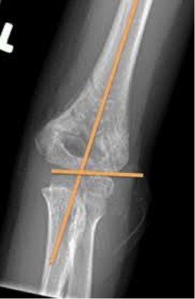
Normal AP flim showing Baumann's angle: angle btw humerus and capitellar physis. This measures amount of varus/valgus deformity Supracondylar Humerus Fractures:
Two categories: · Extension: Distal fragment displaced anteriorly (95% of cases). · Flexion: Distal fragment displaced posteriorly (5% of cases). Four Types: · I: Nondisplaced: look for posterior fat pad · II: Displaced. Posterior cortex intact · III: Completely displaced · IV: Complete periosteal disruption with instability on flexion and extension. Presentation: · Usually from fall on outstretched hand. · Frequently will have neurologic findings: · Anterior Interosseus Neuropraxia: · Most common neurologic finding. Particularly with extension-type fractures. · AIN is a branch of Median nerve. · Patient's cannot flex thumb IP joint or index DIP joint (Can't make an “OK” sign). Almost all will resolve with conservative management. Also have vascular compromise in approximately 1% Usually brachial artery compromise High collateral flow, so patient may have a pink, but pulseless extremity. Still requires emergent reduction. Treatment: Type I: Immobilization at 90 degrees and ortho follow-up. Type II: Closed reduction unless displacement is minimal. Adequate reduction: Baumann's angle wnl, anterior humeral line transects capitellum Type III: High-risk for neurovascular complications. Get ortho involved. Almost always require closed reduction + pinning vs. open reduction Type IV: Open surgical reduction and fixation Indications for open reduction: 1.) Inadequate reduction with closed techniques 2.) Vascular injury 3.) open fracture 4.) Type iv fracture HPI: Patient punched a wall Physical exam: Right hand with significant soft tissue swelling dorsally and TTP over third metacarpal.  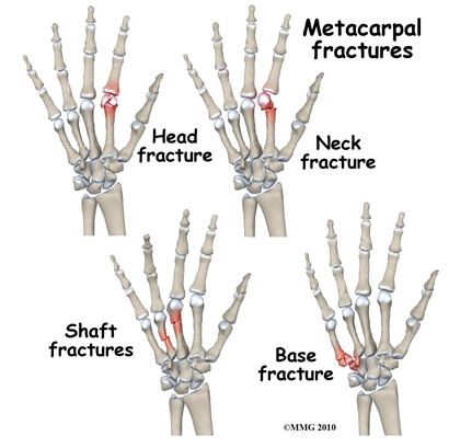 Metacarpal Fractures: General Principles
Acceptable Angulation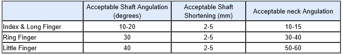 Treatment:
Immobilization: Indications for non-operative immobilization are 1.) Stable fracture pattern 2.) Acceptable angulation 3.) no rotational deformity 4.) Shortening of less than 5mm Splinting: • Fracture splints should be forearm-based and should allow for motion of the interphalangeal (IP) joints. • Splints should extend over the dorsal and palmar aspect of the entire metacarpal being treated. • Generally, the wrist should be placed in 20-30° of extension; the metacarpophalangeal (MCP) joints should be immobilized in 70-90° of flexion, with the dorsal aspect of the splint extending to the IP joints; and the volar aspect should end at the distal palmar crease. • Buddy taping the fingers of the involved metacarpal can aid in maintaining rotational control. HPI: Patient presents with a grossly deformed right upper extremity. PE: Gross deformity of humerus, no open fracture. Unable to extend wrist. Unable to hyper-extend MP joints of fingers and unable to flex IP joint of thumb. Radial and ulnar pulses intact. IMAGING: Multi-factorial fracture along the mid third of humerus along the expected course of the radial nerve. DISPO (if at free standing ED): Transfer patient to ED with on-call orthopedics for definitive fixation. Reduce and splint prior to transfer. TREATMENT: Nonoperative managment. Splinted and cast at bedside. Follow radial nerve palsy clinically for improvement. 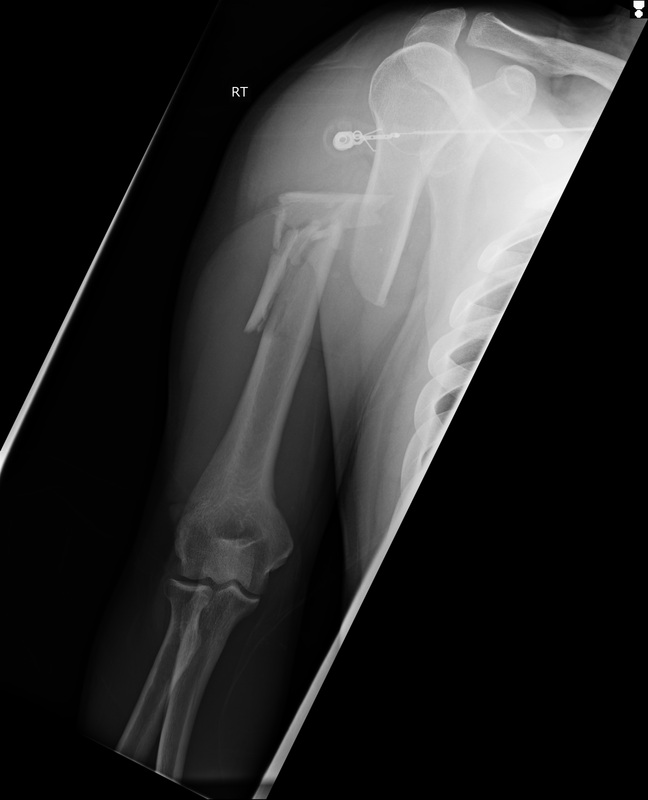 |
Orthopedics Blog
AuthorCMC ER Residents Archives
June 2018
Categories
All
Disclaimer: All images and x-rays included on this blog are the sole property of CMC EM Residency and cannot be used or reproduced without written permission. Patient identifiers have been redacted/changed or patient consent has been obtained. Information contained in this blog is the opinion of the author and application of material contained in this blog is at the discretion of the practitioner to verify for accuracy.
|
 RSS Feed
