CMC COMPENDIUM
  • RESIDENCY
    • About CMC
    • Curriculum
    • Benefits
    • Explore Charlotte
    • Official Site
  • FELLOWSHIP
    • EMS
    • Global EM
    • Pediatric EM
    • Toxicology >
      • Tox Faculty
      • Tox Application
    • (All Others)
  • PEOPLE
    • Program Leadership
    • PGY-3
    • PGY-2
    • PGY-1
    • Alumni
  • STUDENTS/APPLICANTS
    • Medical Students at CMC
    • EM Acting Internship
    • Healthcare Disparities Externship
    • Resident Mentorship
  • #FOAMed
    • EM GuideWire
    • CMC Imaging Mastery
    • Pediatric EM Morsels
    • Blogs, etc. >
      • CMC ECG Masters
      • Core Concepts
      • Cardiology Blog
      • Dr. Patel's Coding Blog
      • Global Health Blog
      • Ortho Blog
      • Pediatric Emergency Medicine
      • Tox Blog
  • Chiefs Corner
    • Top 20
    • Current Chiefs
    • Schedules >
      • Conference/Flashpoint
      • Block Schedule
      • ED Shift Schedule
      • AEC Moonlighting
      • Journal Club/OBP/Audits Schedule
      • Simulation
    • Resources >
      • Fox Reference Library
      • FlashPoint
      • Airway Lecture
      • Student Resources
      • PGY - 1
      • PGY - 2
      • PGY - 3
      • Simulation Reading
      • Resident Wellness
      • Resident Research
      • Resume Builder
    • Individualized Interactive Instruction

Don't  Swing  A  Hammer  At  This  Nail!

5/25/2015

0 Comments

 
HPI:
Right hand dominant middle aged female with history of HTN presents to the ED after accidentally hitting her left index finger with a hammer while working on a home improvement project.   Finger is painful and swollen but she has no other injuries and reports she is otherwise feeling well.

Physical Examination:
Erythematous, swollen, and tender left 2nd distal phalanx. There is a subungual hematoma present over approx. 75% of the nail but the nail is intact.  Motor and sensation intact, full ROM of PIP and DIP, 2+ radial pulse, no other injuries identified.

Radiology:
AP, lateral and oblique views of left 2nd phalanx – no fracture or dislocation identified

Management:
Nail removal and bedside repair of nail bed laceration, tetanus updated, discharged home with 48 hour follow up for reevaluation

Discussion:
Fingertip injuries = most common hand injuries seen in the ED
Most common mechanisms:
  • Crush between two objects
  • Catching finger in a door
  • Power/home tools
  • Direct blows (hammer injury)

Evaluate for:

  • Subungual hematoma
  • Laceration to the nail bed
  • Avulsion of the nail bed
  • DIP fractures or dislocations

Complications of injury:

  • Nail loss
  • Abnormal growth
  • Nonadherence of new nail
  • Splitting of the nail
  • Soft tissue infection
  • Osteomyelitis of the underlying distal tuft
Picture
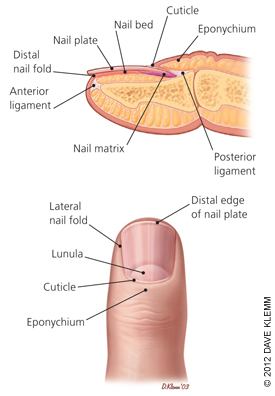
Image obtained from Am Fam Physician. 2012 Apr 15;85(8):779-787.
Fingertip Anatomy:
  • Nail = Desiccated, keratinized squamous cells
  • Perionychium = Nailbed + paronychia
  • Nailbed = Soft tissue beneath the nail that is bound to underlying periosteum of the distal phalanx, consists of the germinal and sterile matrix 
  • Paronychia = Lateral nail folds 
  • Hyponychium  = the skin distal and palmar to the nail – at the junction between the nailbed and fingertip. Contains a lot of PMNs and lymphocytes – helps protect subungal tissue from infection 
  • Nail fold = Holds the proximal nail 
  • Eponychium = aka the cuticle -- or the dorsal, distal portion of the nail fold where it attaches to the dorsum of the nail
  • Lunula = The white part of the proximal nail just distal to the eponychium. Caused by nail cell nuclei in germinal matrix moving upwards and distally to create new nail
  • Sterile Matrix = soft tissue deep to the nail and distal to the lunula, adheres to the nail
  • Germinal Matrix = soft tissue deep to the nail and proximal to the sterile matrix, responsible for most of the nail development; the extensor tendon inserts <1.5mm proximally to this structure

Blood Supply and Innervation:

  • Two volar arterial arches -- anastomoses between digital arteries just above the periosteum of distal phalanx
  • Venous drainage coalesces in proximal nailbed and proximal to nail fold, drains over the dorsum of the finger
  • Perionychium innervated by dorsal branches of the paired digital nerves

Nail Growth:

  • Longitudinal nail growth = 70-160 days to cover entire length
  • After an injury, growth may be stunted/absent for up to 21 days, then nail grows rapidly for approx. 50 days then resumes normal growth rate
  • Results in a characteristic “lump” that often forms after traumatic injury

Treatment:

Obtain radiographs to rule out distal phalanx fracture
Evaluate for subunginal hematoma and nail bed lacerations

Drain hematoma if <50% nail involved 
  • Puncture with sterile needle vs electrocautery 

Nail removal, I&D, repair of nail bed if >50% of nail involved

  • Remove nail
  • Repair nail bed with 6-0 absorbable suture or Dermabond
  • Splint eponychial fold with original nail (if possible), aluminum splint, or non-adherent gauze to prevent scarring
0 Comments

Patella  Fractures

5/18/2015

0 Comments

 
HPI:
20ish year old and otherwise healthy male presents as the restrained driver in head-on MVC at low speed just prior to arrival. He has a large linear laceration to anterior aspect of R knee and is unable to fully extend the knee. Wound is hemostatic. No other injuries or complaints.

Physical Examination:
10cm linear, horizontal laceration to the anterior aspect of the patient’s R knee.  Knee is held in slightly flexed positioning. Decreased ROM of the knee secondary to pain and injury, patient unable to actively extend extremity in straight leg raise.  FROM of ankle. 2+ DP and PT pulses present. Sensation intact to light touch throughout. No erythema, swelling or significant tenderness present over RLE. 

Radiology:

AP/Lateral/Sunrise/Oblique views of R knee:
Transverse fracture of the patella with 3mm of displacement present and surrounding soft tissue edema

Management:
  • Bedside washout of wound with copious saline and with wet gauze placement
  • Tetanus updated
  • 2g IV Ancef administered
  • Admission for ORIF of R patella with tension band construction

Discussion:
  • Patella = largest sesamoid bone in the body with the thickest articular cartilage 
  • Patella fractures = 1% of all bony injuries
  • Injury usually by direct impact or indirect eccentric contraction of quadriceps with hyperextension of the knee
  • Usually seen in younger adult population (20-50 years old)

 Types of fracture:
Picture
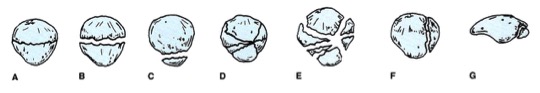
Figure obtained from jaaos.org; Patellar Fractures in Adults; AAOS April 2011 vol. 19 no. 4 198-207,Figure 4
A)  Nondisplaced
B)  Transverse
C)  Pole or sleeve (upper or lower) 
D)  Comminuted nondisplaced
E)   Comminuted displaced
F)   Vertical
G)  Osteochondral

Evaluate with physical examination first
Severe knee pain and soft tissue swelling should raise suspicion for injury
Concerning findings:
  • Palpable bony defect
  • Large hemarthrosis
  • High riding patella
  • Inability to weight bear
  • Active extension of knee difficult to perform
  • Unable to perform straight leg raise test = retinaculum disrupted (the connective tissue band around tendons that hold them in place)
Picture
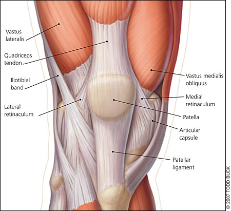
Image obtained from aafp.org, Am Fam Physician. 2007 Jan 15;75(2):194-202.
Obtain Radiographs
Fracture displacement best viewed on lateral films, but obtain AP/Lat/Oblique/Sunrise to fully evaluate
Degree of displacement =  degree of retinacular disruption

Treatment:
Nonoperative:
  • If intact extensor mechanism
  • Nondisplaced or minimally displaced fracture
  • Vertical fracture
Knee immobilized in extension with brace or cylinder cast with early WBAT
Early active ROM with hinged knee brace after 2-3 weeks

Operative:
  • Open fractures
  • Extensor mechanism failure
  • Fracture with articular displacement of >2mm
  • Displaced patella fracture >3mm
  • Patella sleeve fractures in children
Usually managed by ORIF with tension band construction

Pearls:
Watch out for patella sleeve fractures in peds – need high index of suspicion to diagnose
  • Small avulsion fractures that occur between cartilage “sleeve” and main bony patella usually in kids 8-12 yrs old when patellar ossification is nearly complete
Consider obtaining MRI in a child with normal x-rays but who is unable to straight leg raise

Bipartite patella = failure of bony fusion
  • Anatomic anomaly seen in up to 8% of population and may be mistaken for fracture
  • May be bilateral in up to 50% of people with this variation

Any lacerations present must be assumed to communicate with the knee joint until this assumption is disproved by a saline load joint challenge

A patellar fracture is problematic and requires intervention if extensor mechanism of knee is nonfunctional = unable to perform straight leg raise test
0 Comments

Who  let  the  dogs  out?  - Pyogenic  Flexor Tenosynovitis

5/10/2015

0 Comments

 
HPI:
Pre-teen male with no PMH presents to the ED after a dog bite the night before. Patient was bit on the palm of his left hand by a relative’s pit bull known to be fully vaccinated. Wound had been becoming more painful and 3rd digit is more swollen, painful, and difficult to range. No other injuries or complaints.

Physical Exam:
Two sub-centimeter puncture wounds to the center of the palm of the left hand, hemostatic with small amount of surrounding erythema present. 3rd digit with fusiform soft tissue swelling, increased warmth, and significant tenderness to palpation. Finger in slightly flexed positioning. Decreased ROM of the 3rd digit with flexion and significant pain with passive extension. Sensation intact to light touch throughout digits and hand. No other erythema, swelling, or significant tenderness present over LUE.  2+ palpable radial and ulnar pulses present.

Radiology:
No fracture or acute changes. No foreign body present.

Management:
Patient was admitted and was started on IV antibiotics for suspected pyogenic flexor tenosynovitis. Wound was rechecked in the morning with minimal improvement in symptoms and patient underwent I&D of digit and hand with continued IV antibiotics.

Discussion:
Pyogenic flexor tenosynovitis = infection of synovial sheath surrounding the flexor tendon
  • Staph aureus = most common
  • Eikenella = human bites
  • Pasteurella multocida = animal bites

Pain/swelling usually delayed 24-48 hours, usually localized to the palmar aspect of one digit of the hand

Kanavel signs – key physical exam findings for diagnosis
  • Flexed posturing of the digit
  • Tenderness to palpation over tendon sheath
  • Significant pain with passive extension of digit
  • Fusiform swelling of the digit


Obtain x-rays to rule out bony involvement or foreign body

MRI can help diagnose but is expensive and generally unnecessary since clinical exam usually is sufficient

Treatment:
If early presentation:   
  • Admission
  • IV abx
  • Immobilization
  • Observation

If late presentation or if no improvement after 24 hours of conservative treatment:
  • Operative I&D with IV antibiotics

Empiric Abx to consider:
Otherwise healthy individuals--
  • Cefazolin 1-2 g IV q6-8h
  • Clindamycin 600 mg IV q8h 
  • Erythromycin 500-1000 mg IV q6h 
Immunocompromised individuals or bite involvement:
  • Ampicillin-sulbactam 1.5-3 g IV q6h
  • Cefoxitin 2 g IV q6-8h
  • Clindamycin 600 mg IV q8h PLUS levofloxacin 500 mg IV q24h 

 Consider with dog bites-- most are polymicrobial:
  • Staph
  • Strep
  • Pasteurella multocida
  • Anerobes

Key Points:

History and physical examination is key to diagnosis
Start antibiotics early 
Consider surgical intervention when there is:
  • crushed tissue
  • bite to pulp space
  • involvement of:
                o   nail bed 
                o   flexor tendon sheath
                o   deep spaces of the palm
                o   joint spaces

Kanavel signs may not be seen if patient has:
  • Early presentation
  • Antibiotics recently administered
  • Immunocompromised state/Chronic infection/Diabetes
0 Comments

    Orthopedics Blog

    Picture

    Author 

    CMC ER Residents 
    (for learning purposes, imagine all cases present to a free standing ED for dispo and treatment options)

    Subscribe To Blog

    Archives

    June 2018
    April 2018
    March 2018
    October 2017
    September 2017
    February 2016
    January 2016
    December 2015
    November 2015
    October 2015
    September 2015
    August 2015
    July 2015
    June 2015
    May 2015
    April 2015
    March 2015
    February 2015
    January 2015
    December 2014
    November 2014

    Categories

    All
    Lower Extremity
    Nerve Injury
    Spine
    Sports Medicine
    Upper Extremity
    Vascular Injury

    RSS Feed

    Disclaimer: All images and x-rays included on this blog are the sole property of CMC EM Residency and cannot be used or reproduced without written permission.  Patient identifiers have been redacted/changed or patient consent has been obtained.  Information contained in this blog is the opinion of the author and application of material contained in this blog is at the discretion of the practitioner to verify for accuracy.
Powered by Create your own unique website with customizable templates.
  • RESIDENCY
    • About CMC
    • Curriculum
    • Benefits
    • Explore Charlotte
    • Official Site
  • FELLOWSHIP
    • EMS
    • Global EM
    • Pediatric EM
    • Toxicology >
      • Tox Faculty
      • Tox Application
    • (All Others)
  • PEOPLE
    • Program Leadership
    • PGY-3
    • PGY-2
    • PGY-1
    • Alumni
  • STUDENTS/APPLICANTS
    • Medical Students at CMC
    • EM Acting Internship
    • Healthcare Disparities Externship
    • Resident Mentorship
  • #FOAMed
    • EM GuideWire
    • CMC Imaging Mastery
    • Pediatric EM Morsels
    • Blogs, etc. >
      • CMC ECG Masters
      • Core Concepts
      • Cardiology Blog
      • Dr. Patel's Coding Blog
      • Global Health Blog
      • Ortho Blog
      • Pediatric Emergency Medicine
      • Tox Blog
  • Chiefs Corner
    • Top 20
    • Current Chiefs
    • Schedules >
      • Conference/Flashpoint
      • Block Schedule
      • ED Shift Schedule
      • AEC Moonlighting
      • Journal Club/OBP/Audits Schedule
      • Simulation
    • Resources >
      • Fox Reference Library
      • FlashPoint
      • Airway Lecture
      • Student Resources
      • PGY - 1
      • PGY - 2
      • PGY - 3
      • Simulation Reading
      • Resident Wellness
      • Resident Research
      • Resume Builder
    • Individualized Interactive Instruction