CMC COMPENDIUM
  • RESIDENCY
    • About CMC
    • Curriculum
    • Benefits
    • Explore Charlotte
    • Official Site
  • FELLOWSHIP
    • EMS
    • Global EM
    • Pediatric EM
    • Toxicology >
      • Tox Faculty
      • Tox Application
    • (All Others)
  • PEOPLE
    • Program Leadership
    • PGY-3
    • PGY-2
    • PGY-1
    • Alumni
  • STUDENTS/APPLICANTS
    • Medical Students at CMC
    • EM Acting Internship
    • Healthcare Disparities Externship
    • Resident Mentorship
  • #FOAMed
    • EM GuideWire
    • CMC Imaging Mastery
    • Pediatric EM Morsels
    • Blogs, etc. >
      • CMC ECG Masters
      • Core Concepts
      • Cardiology Blog
      • Dr. Patel's Coding Blog
      • Global Health Blog
      • Ortho Blog
      • Pediatric Emergency Medicine
      • Tox Blog
  • Chiefs Corner
    • Top 20
    • Current Chiefs
    • Schedules >
      • Conference/Flashpoint
      • Block Schedule
      • ED Shift Schedule
      • AEC Moonlighting
      • Journal Club/OBP/Audits Schedule
      • Simulation
    • Resources >
      • Fox Reference Library
      • FlashPoint
      • Airway Lecture
      • Student Resources
      • PGY - 1
      • PGY - 2
      • PGY - 3
      • Simulation Reading
      • Resident Wellness
      • Resident Research
      • Resume Builder
    • Individualized Interactive Instruction

Knee Dislocation

10/17/2015

0 Comments

 
HPI: 
Otherwise healthy 27-year-old male who presents with right knee pain.  The patient is a construction worker and while on a construction site today, a wooden wall fell onto him and other workers.  The patient received an isolated injury to his right knee and presents with pain and deformity.  Patient denies any other injuries, his vital signs are within normal limits.

​Exam:
Patient lying in a supine position.  Right knee has no significant effusion, erythema, or overlying abrasions/lacerations.  Palpation of the right knee reveals a large step off between the patella and anterior surface of the femur.  Patient is neurovascularly intact distally.  Representative x-ray is shown below.
Picture
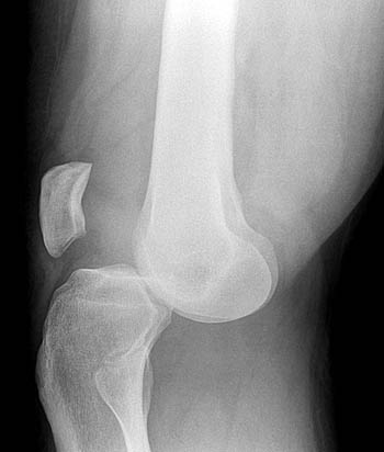
http://radsource.us/
 
Discussion:
There are 5 different types of knee dislocations based on the direction of displacement from the tibia.
  1. Anterior is the most common.  This is due to hyperextension injury and is often associated with a tear of the posterior cruciate ligament.  Arterial injury is usually secondary to intimal tear.
  2. Posterior is the second most common type.  This is usually due to an axial load to a flexed knee, such as in a MVC with dashboard injury.  This has the highest rate of associated popliteal artery injuries.
  3. Lateral dislocation is usually due to varus or valgus force.  These typically involve tears of the anterior cruciate ligament and posterior cruciate ligament.  These are associated with peroneal nerve injury.
  4. Medial dislocation is usually due to varus or valgus force.  This is usually associated with posterior cruciate ligament injury.
  5. Rotational dislocation is commonly associated with sports injuries.  Posterio-lateral is the most common rotation direction.

​Physical exam often times result with no obvious deformity.  It has been estimated that as many as 50% spontaneously reduce prior to arrival to the ED.  In the emergency department, vascular examination is of utmost importance.  With obvious deformity and absent pulses, reduction must take place immediately, before radiographs are obtained.  After reduction, dorsalis pedis and posterior tibial pulses should be palpated, but the palpation of pulses does not rule out arterial injury.  The next step should be measuring ankle-brachial index of the affected side.  If the ABI is normal (> 0.9), then you can monitor the patient with repeat examinations as this has a 100% negative predictive value for arterial injury.  If the ABI is abnormal (<0.9), then a CT angiography should be performed along with vascular surgery consultation.  If pulses are still not palpable after successful reduction, immediate surgical exploration should be performed.

There is one situation where reduction of the knee should not take place.  The “dimple sign” represents buttonholing of the medial femoral condyle through the anterior medial joint capsule.  The sign indicates an irreducible dislocation and closed reduction is contraindicated for the risk of skin necrosis.

Picture
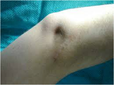
http://jbjs.org/content/87/7/1571

Initial imaging consists of plain radiographs.  Remember, many of these reduce prior to arrival and the radiograph may appear normal.  Despite this, you should look closely for avulsion fractures and osteochondral defects.  CT angiography should be obtained as discussed above if necessary.
After reduction and verification of ABI, the patient should be placed in a splint with 20-30° of flexion.  Post reduction films should be taken after the splint or brace is applied to confirm placement.  If emergent surgical intervention is not warranted, many of these require delayed ligamentous repair.
Long-term complications consist of arthritic symptoms, instability of the joint, peroneal nerve injury, and chronic vascular compromise.

Management Pearls:
  • Deformity with no pulses = immediate reduction
  • Return of pulses after reduction does not rule out arterial injury
  • After reduction, obtain ABI.  Normal ABI has 100% negative predictive value of arterial injury
  • If abnormal ABI, proceed with CT angiography and vascular consult
  • Knee should be splinted in 20-30° of flexion
  • Patient will likely require surgical repair of ligamental injury

​References:
1.  Patel, Anay.  "Knee Dislocation."  Orthobullets 08/11/15.  Retrieved 10/15/15 from http://www.orthobullets.com/trauma/1043/knee-dislocation.
2.  Peskun, C.J. et al.  "Diagnosis and Management of Knee Dislocations."  The Physician and Sportsmedicine.  Dec, 2010.  4:38, pages  101-111.

By Dr. Phillip Lunsford
0 Comments

    Orthopedics Blog

    Picture

    Author 

    CMC ER Residents 
    (for learning purposes, imagine all cases present to a free standing ED for dispo and treatment options)

    Subscribe To Blog

    Archives

    June 2018
    April 2018
    March 2018
    October 2017
    September 2017
    February 2016
    January 2016
    December 2015
    November 2015
    October 2015
    September 2015
    August 2015
    July 2015
    June 2015
    May 2015
    April 2015
    March 2015
    February 2015
    January 2015
    December 2014
    November 2014

    Categories

    All
    Lower Extremity
    Nerve Injury
    Spine
    Sports Medicine
    Upper Extremity
    Vascular Injury

    RSS Feed

    Disclaimer: All images and x-rays included on this blog are the sole property of CMC EM Residency and cannot be used or reproduced without written permission.  Patient identifiers have been redacted/changed or patient consent has been obtained.  Information contained in this blog is the opinion of the author and application of material contained in this blog is at the discretion of the practitioner to verify for accuracy.
Powered by Create your own unique website with customizable templates.
  • RESIDENCY
    • About CMC
    • Curriculum
    • Benefits
    • Explore Charlotte
    • Official Site
  • FELLOWSHIP
    • EMS
    • Global EM
    • Pediatric EM
    • Toxicology >
      • Tox Faculty
      • Tox Application
    • (All Others)
  • PEOPLE
    • Program Leadership
    • PGY-3
    • PGY-2
    • PGY-1
    • Alumni
  • STUDENTS/APPLICANTS
    • Medical Students at CMC
    • EM Acting Internship
    • Healthcare Disparities Externship
    • Resident Mentorship
  • #FOAMed
    • EM GuideWire
    • CMC Imaging Mastery
    • Pediatric EM Morsels
    • Blogs, etc. >
      • CMC ECG Masters
      • Core Concepts
      • Cardiology Blog
      • Dr. Patel's Coding Blog
      • Global Health Blog
      • Ortho Blog
      • Pediatric Emergency Medicine
      • Tox Blog
  • Chiefs Corner
    • Top 20
    • Current Chiefs
    • Schedules >
      • Conference/Flashpoint
      • Block Schedule
      • ED Shift Schedule
      • AEC Moonlighting
      • Journal Club/OBP/Audits Schedule
      • Simulation
    • Resources >
      • Fox Reference Library
      • FlashPoint
      • Airway Lecture
      • Student Resources
      • PGY - 1
      • PGY - 2
      • PGY - 3
      • Simulation Reading
      • Resident Wellness
      • Resident Research
      • Resume Builder
    • Individualized Interactive Instruction