|
HPI: Middle-aged male s/p MCC. PE: Ecchymoses, swelling, and tenderness about ankle. Tender over proximal fibula and pain with squeezing of calf. Figure 1: Mortise view of ankle showing widened mortise medially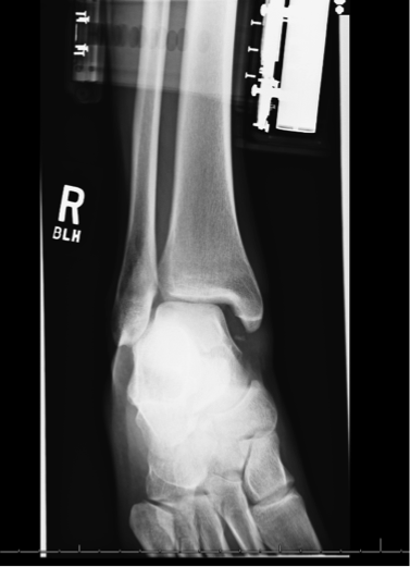 Figure 2: Leg film showing proximal fibular fracture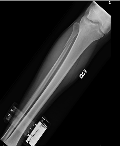 Ankle Sprains: - Divided into grades o I: Minor, no significant ligamentous damage. Able to bear weight. o II: Associated with partial ligamentous tear. Significant ecchymoses/swelling. Difficulty bearing weight. o III: Associated with complete ligamentous tear. Significant functional loss and universal inability to bear weight. - When should we obtain radiographs?: o If pretest clinical suspicion is high based on mechanism of injury or patient cannot be ruled out for fracture based on Ottawa ankle rules: 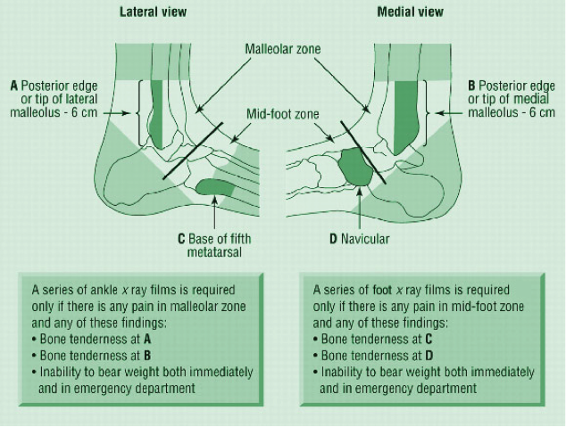 Syndesmotic injuries (AKA “high” ankle sprains):
o PE and mechanism:
o Treatment:
- Otherwise patient needs syndesmotic screw fixation. Patient can be immobilized and follow-up as an outpatient for surgical fixation
0 Comments
HPI: 7 y/o fall from monkey bars. Landed on extended shoulder + outstretched arm. Physical exam: Obvious arm deformity. Ecchymosis over distal/medial arm. Inability to flex thumb IP joint and DIP of index finger (AIN neuropraxia). Palpable pulses. Warm extremity. 
AP showing mild varus angulation 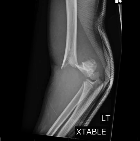
Lateral film showing significant posterior displacement of distal portion of fracture 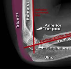
Normal lateral film 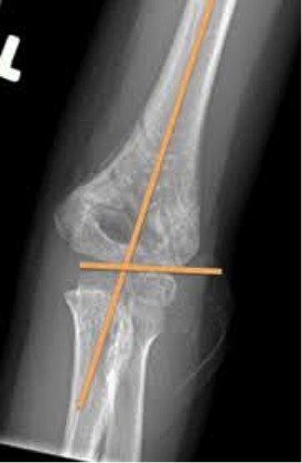
Normal AP flim showing Baumann's angle: angle btw humerus and capitellar physis. This measures amount of varus/valgus deformity Supracondylar Humerus Fractures:
Two categories: · Extension: Distal fragment displaced anteriorly (95% of cases). · Flexion: Distal fragment displaced posteriorly (5% of cases). Four Types: · I: Nondisplaced: look for posterior fat pad · II: Displaced. Posterior cortex intact · III: Completely displaced · IV: Complete periosteal disruption with instability on flexion and extension. Presentation: · Usually from fall on outstretched hand. · Frequently will have neurologic findings: · Anterior Interosseus Neuropraxia: · Most common neurologic finding. Particularly with extension-type fractures. · AIN is a branch of Median nerve. · Patient's cannot flex thumb IP joint or index DIP joint (Can't make an “OK” sign). Almost all will resolve with conservative management. Also have vascular compromise in approximately 1% Usually brachial artery compromise High collateral flow, so patient may have a pink, but pulseless extremity. Still requires emergent reduction. Treatment: Type I: Immobilization at 90 degrees and ortho follow-up. Type II: Closed reduction unless displacement is minimal. Adequate reduction: Baumann's angle wnl, anterior humeral line transects capitellum Type III: High-risk for neurovascular complications. Get ortho involved. Almost always require closed reduction + pinning vs. open reduction Type IV: Open surgical reduction and fixation Indications for open reduction: 1.) Inadequate reduction with closed techniques 2.) Vascular injury 3.) open fracture 4.) Type iv fracture HPI: Patient punched a wall Physical exam: Right hand with significant soft tissue swelling dorsally and TTP over third metacarpal. 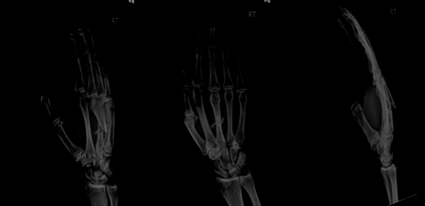 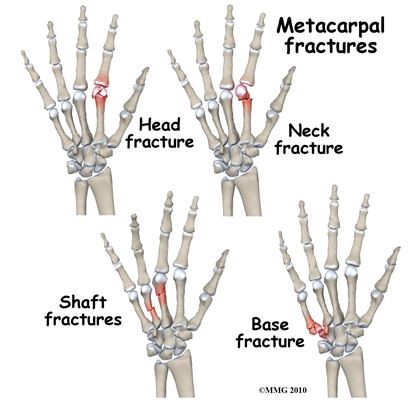 Metacarpal Fractures: General Principles
Acceptable Angulation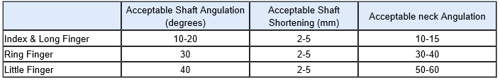 Treatment:
Immobilization: Indications for non-operative immobilization are 1.) Stable fracture pattern 2.) Acceptable angulation 3.) no rotational deformity 4.) Shortening of less than 5mm Splinting: • Fracture splints should be forearm-based and should allow for motion of the interphalangeal (IP) joints. • Splints should extend over the dorsal and palmar aspect of the entire metacarpal being treated. • Generally, the wrist should be placed in 20-30° of extension; the metacarpophalangeal (MCP) joints should be immobilized in 70-90° of flexion, with the dorsal aspect of the splint extending to the IP joints; and the volar aspect should end at the distal palmar crease. • Buddy taping the fingers of the involved metacarpal can aid in maintaining rotational control. |
Orthopedics Blog
AuthorCMC ER Residents Archives
June 2018
Categories
All
Disclaimer: All images and x-rays included on this blog are the sole property of CMC EM Residency and cannot be used or reproduced without written permission. Patient identifiers have been redacted/changed or patient consent has been obtained. Information contained in this blog is the opinion of the author and application of material contained in this blog is at the discretion of the practitioner to verify for accuracy.
|
 RSS Feed
