|
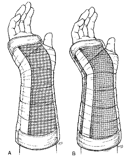
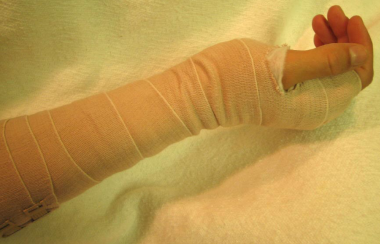
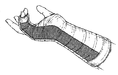
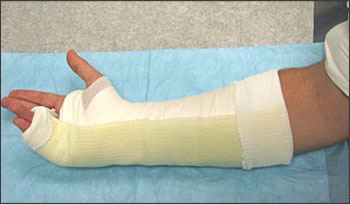
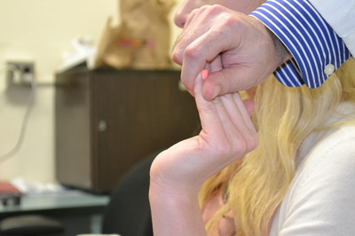
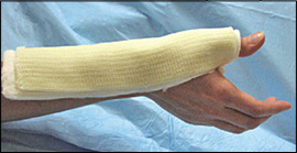
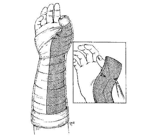
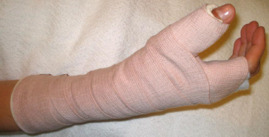
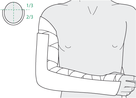
Volar Splint Indications: -Hand and Wrist injures (NOT distal radius or ulna fractures, can still supinate and pronate) -Carpal fractures -Lunate dislocation -2nd-5th metacarpal head fracture Application: -Extends along volar forearm from metacarpal heads to just proximal to radial head -Allow flexion of elbow -Wrist at 20 degrees of extension -Can add dorsal "sandwich" for stability Ulnar Gutter Splint Indications: -4th and 5th phalanges and metacarpals Application: -Extends from 5th DIP to proximal forearm -Wrist at 20 degrees of extension -Flex MCPs at 50-70 degrees, PIP and DIPs in slight flexion Thumb Spica Splint Indications: -Scaphoid and lunate fractures -1st metacarpal fracture -Thumb fractures -De Quervain tenosynovitis Application: -Extends from tip of thumb to proximal forearm -Wrist at 20 degrees of extension -Thumb slightly flexed Long Arm Splint Indications: -Proximal forearm and elbow fractures -Intraarticular fractures of distal humerus and olecranon Application: -Elbow at 90 degrees of flexion -Neutral forearm and wrist Sugar Tong Splint Indications: -Wrist and distal forearm fractures Application: -Extends from MCPs on dorsum, around elbow, to volar midpalmar crease -Elbow at 90 degrees of flexion -Neutral forearm and wrist -Double sugar tong for complex or unstable forearm and elbow fractures Resources:
Michael T Fitch, MD. Basic Splinting Techniques. New England Journal of Medicine. 2008; 359:e32. Wikiem.com Ortho-teaching.feinberg.northwestern.edu/docs/Splinting.pptx
0 Comments
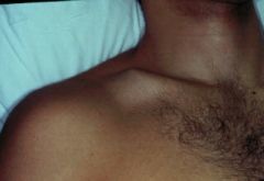
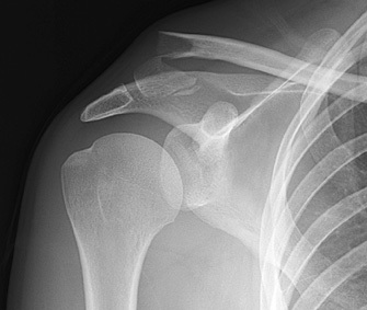
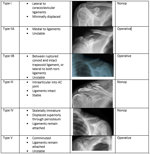
HPI: 23 yo male s/p MCC. Patient reports that he swerved to avoid hitting a vehicle in front of him that stopped abruptly and layed down his bike, landing on his right shoulder. He was helmeted and did not lose consciousness. Ambulatory after the event, hemodynamically stable, and complaining of right shoulder pain. Exam: Radiology: Management: Middle Third (80-85%) Lateral Third (10-15%) Medial Third (5-8%) Treatment:
Nonop
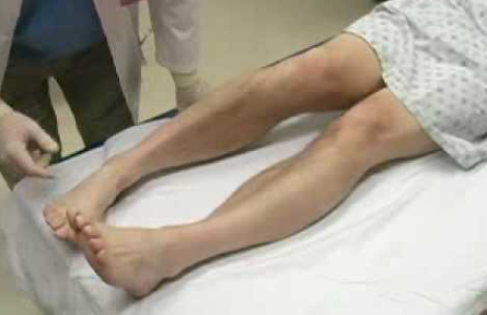
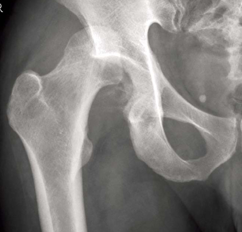
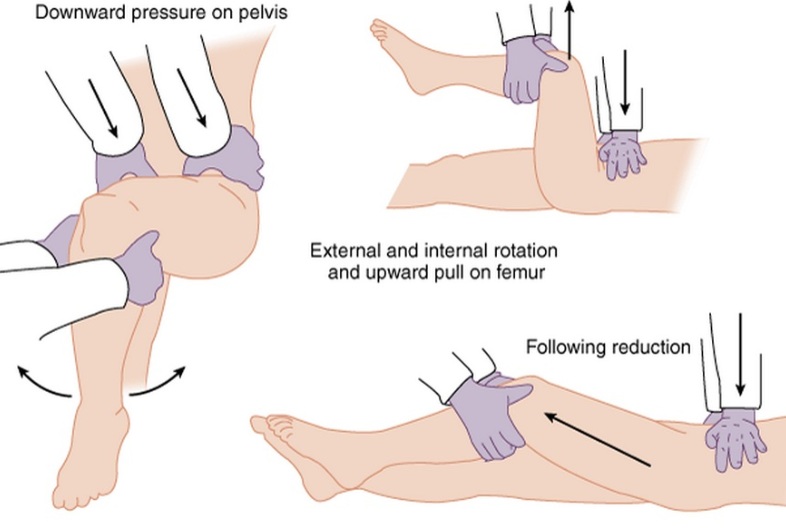
Resources: Orthobullets.com HPI: 22 yo otherwise healthy male presents s/p head on MVC vs tree. Patient is awake and alert, hemodynamically stable, complaining of right hip pain. Physical Exam: No external signs of trauma. Right lower extremity is shortened compared to the left and internal rotated. No numbness, 2+ DP pulse. Classification: - Simple: pure dislocation - Complex: with associated fracture of acetabulum or proximal femur Mechanism: - Axial load on femur while hip flexed and adducted or through flexed knee (dashboard injury such as this patient) Requires emergent reduction (within 6 hours!) due to risk of vascular compromise to hip and osteonecrosis However... Examine femoral neck closely on XR to rule out fracture prior to attempting closed reduction. With ipsilateral femoral neck fracture, closed reduction is contraindicated! Patient must be adequately sedated for procedure. Propofol helps with tissue relaxation! Post reduction CT must be performed to evaluate for: - femoral head fractures - loose bodies - acetabular fractures Commonly associated with ipsilateral knee injuries (up to 25%) Dispo: For simple dislocation, protected weight bearing for 4-6 weeks Resources: 1. Serna, Fernando MD, Corczyca, John MD. Hip Dislocations and Femoral Head Fractures. University of Rochester Medical Center. March 2004. 2. Orthobullets.com |
Orthopedics BlogAuthorCMC ER Residents Archives
June 2018
Categories
All
Disclaimer: All images and x-rays included on this blog are the sole property of CMC EM Residency and cannot be used or reproduced without written permission. Patient identifiers have been redacted/changed or patient consent has been obtained. Information contained in this blog is the opinion of the author and application of material contained in this blog is at the discretion of the practitioner to verify for accuracy.
|




 RSS Feed
