CMC COMPENDIUM
  • RESIDENCY
    • About CMC
    • Curriculum
    • Benefits
    • Explore Charlotte
    • Official Site
  • FELLOWSHIP
    • EMS
    • Global EM
    • Pediatric EM
    • Toxicology >
      • Tox Faculty
      • Tox Application
    • (All Others)
  • PEOPLE
    • Program Leadership
    • PGY-3
    • PGY-2
    • PGY-1
    • Alumni
  • STUDENTS/APPLICANTS
    • Medical Students at CMC
    • EM Acting Internship
    • Healthcare Disparities Externship
    • Resident Mentorship
  • #FOAMed
    • EM GuideWire
    • CMC Imaging Mastery
    • Pediatric EM Morsels
    • Blogs, etc. >
      • CMC ECG Masters
      • Core Concepts
      • Cardiology Blog
      • Dr. Patel's Coding Blog
      • Global Health Blog
      • Ortho Blog
      • Pediatric Emergency Medicine
      • Tox Blog
  • Chiefs Corner
    • Top 20
    • Current Chiefs
    • Schedules >
      • Conference/Flashpoint
      • Block Schedule
      • ED Shift Schedule
      • AEC Moonlighting
      • Journal Club/OBP/Audits Schedule
      • Simulation
    • Resources >
      • Fox Reference Library
      • FlashPoint
      • Airway Lecture
      • Student Resources
      • PGY - 1
      • PGY - 2
      • PGY - 3
      • Simulation Reading
      • Resident Wellness
      • Resident Research
      • Resume Builder
    • Individualized Interactive Instruction

The Wrist

3/5/2018

0 Comments

 
Authored by Dr. Javier Andrade
  • With any orthopedic injury, it is important to understand the normal bony anatomy of the injured area you are examining:
    • The Wrist is a complex area with multiples bones and joint.
    • It is composed of the more proximal ulna and radius which anchor the wrist and help create the supination and pronation needed for complex hand movements
    • The distal portion of the wrist is made up of the 8 carpal bones arranged in two rows.
Picture
Picture
  • Now that we now what bones make up the wrist, we can talk a bit more about the alignment and tricks we can use to see if there is an injury to the wrist.
    • One such trick we can use for the PA view are Gilula’s Line. It is a technique which uses 3 curved lines to check the alignment of the carpal bone rows.
      • Proximal row, proximal portion (pink)
      • Proximal row, distal portion (blue)
      • Distal row, proximal portion (red)
Picture
  • Also, as with any ortho injury, remember to assess that the patient is neurovascularly intact!
    • Vascular: Can use the doppler probe to assess
      • Ulnar artery
      • Radial Artery
      • Palmar arch
    • Nerves:
      • Radial n: Test the extensor pollicus longus using “thumbs up”
      • Median n: Test using the “OK sign”
        • Make sure patient flexes at the DIP joint
      • Ulnar n: Test using pinky abduction
  • Pathology:
    • Scaphoid Fx
      • Most commonly injured carpal bone and can be easily missed. If you have clinical exam findings but no radiologic findings, treat this as a fracture!
      • Tx: Thumb spica cast and reimage in 12 days.
    • Lunate and Perilunate dislocation
      • Associated with high-energy mechanism of injury as it has to tear through multiple ligaments that hold the hand together. Be on the lookout for other injuries in the wrist.
        • Associated with median n injury in 25% of patients
      • Notice the anatomy on the normal image (left). We can use the lines on the lateral XR view to assess for injury
Picture
Picture
  • Hamate fracture:
    • Body: Clenched fist against solid object
      • Typically requires surgical intervention
    • Hook: Associated with injuries that require swinging equipment (baseball, tennis, etc.)
      • Can cause ulnar n. injury or impingement
  • Distal Radius Fractures:
    • Colle’s: Fx with posterior displacement
    • Smith’s: Fx with anterior displacement
  • Distal Radial-Ulnar joint (DRUJ) injuries
    • Normally, the ulna sits on a small groove on the distal radius known as the sigmoid notch.
      • Any spacing here is abnormal
Picture
  • Management:
    • If purely ligamentous closed immobilization and outpatient follow-up
    • If associated with fracture then operative repair is necessary
0 Comments

Evaluation of the pediatric limp

3/4/2018

0 Comments

 
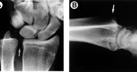
Authored by Dr. Jessica Hoglund
1.   Differential (categorized based on gait pattern)
​A.     Antalgic gait
Picture
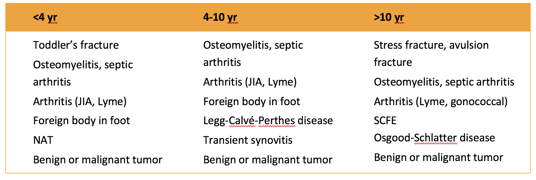
B.     Non-antalgic gait
Picture
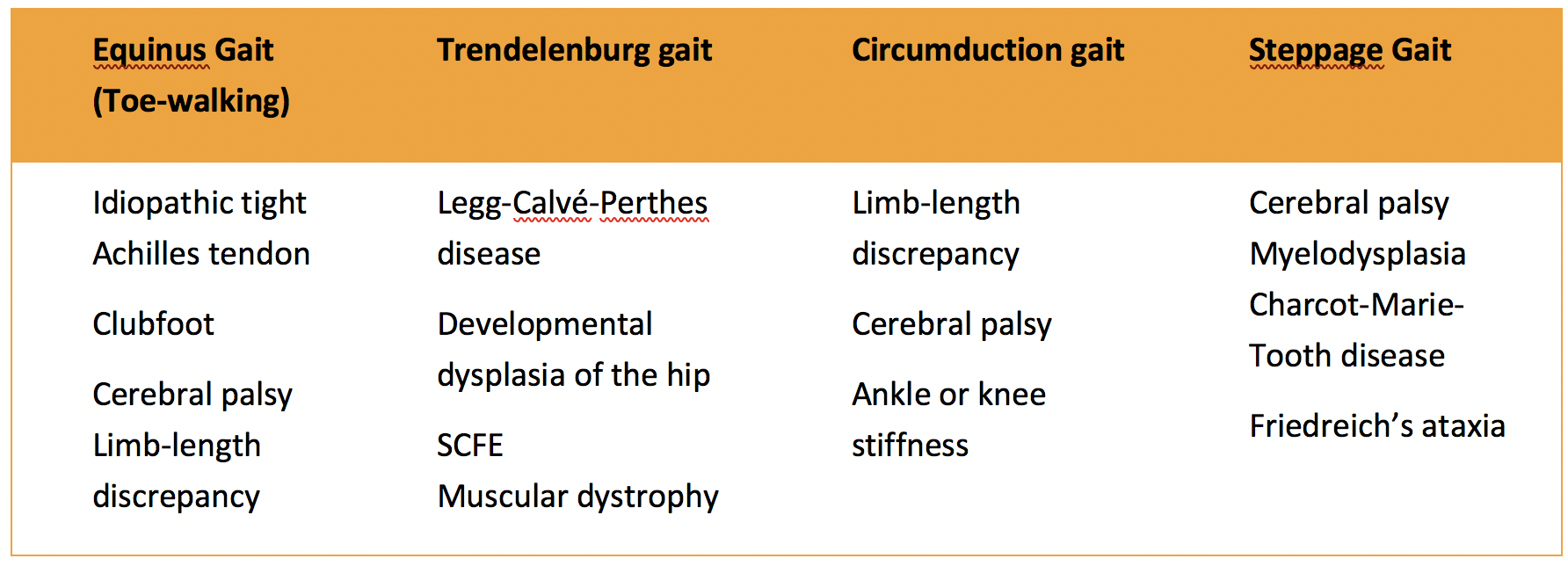
1.   Transient synovitis
A.     Most common cause of hip pain/limp in children <10 yrs
B.     Aseptic inflammation of the hip (postviral etiology)
C.     Diagnosis
        i.  Physical exam:
            1.     Non-toxic
            2.     Antalgic gait    
                   i.         Imaging performed when considering other differentials   
                  ii.         Ultrasound may be performed to confirm the presence of a joint effusion
D.     Management      
        i.         Rest    
​        ii.         Analgesics 

2.   Septic arthritis
A.     Causes      
        i.         Bacterial infection of joint    
        ii.         Most commonly s. aureus   
        iii.         Consider E. coli or GBS in children < 2 months   
        iv.         Consider gonococcal arthritis in adolescents, sexually active patients, and in polyarticular arthritis    
        v.         Consider salmonella in sickle cell disease
B.     Clinical features      
        i.         Neonates often do not appear ill and may not have fever in 50% of cases   
        ii.         Older infants, toddlers, children may localize, limp, or refuse to walk
C.     Diagnosis     
        i.         Physical exam1.     Limb externally rotated, flexed, and abducted    
        ii.         Labs1.     CBC, ESR, possibly blood cultures   
       iii.         Kocher criteria (septic arthritis vs transient synovitis, >3 predictors high risk for septic arthritis)
                  1.     Non weight-bearing
                  2.     Temp > 101.3oF (38.5oC)
                  3.     ESR > 40 mm/hr
                  4.     WBC > 12,000 cells/mm3   
        iv.         Imaging
Picture
Picture
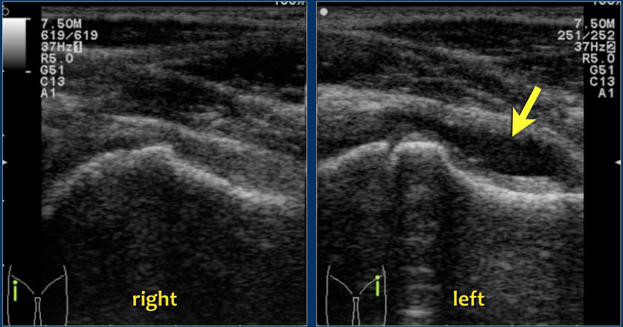
​          v.        Arthrocentesis
                    1.     Ultrasound guided or fluoro guided for hip
                    2.     Synovial fluid with >50,000 WBC’s, >75% PMN’s, positive gram stain and culture
D.     Management      
          i.         IV antibiotics (vanc, rocephin or cefoxatime if < 2 months)    
          ii.         Surgical drainage
E.     Complications      
         i.         Avascular necrosis    
         ii.         Capsule damage   
         iii.         Chronic arthritis   
         iv.         Osteomyelitis    
​         v.         Sepsis
​
​4.   SCFE
A.     MCC of hip disability in adolescents
B.     Obese adolescents whose hips are exposed to repetitive minimal trauma
C.     Diagnosis      
         i.         Physical exam
                   1.     Decreased internal rotation
                   2.    Antalgic or Trendelenburg gait
                   3.    Determine if pt is able to ambulate with or without crutches (determines prognosis)    
        ii.         Imaging
                   1.     Any adolescent with chronic groin, hip, thigh, or knee pain deserves bilateral hip XR (AP and lateral)
                   2.     High incidence of bilateral disease
                  3.     Use Klein’s lines to aid in diagnosis.
​A and C below are abnormal. B and D are normal (line drawn from   superior aspect of femoral neck transects lateral aspect of femoral head).
Picture
Picture
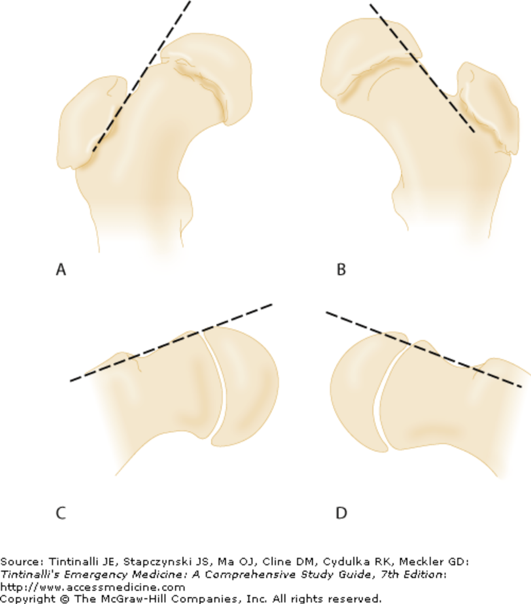
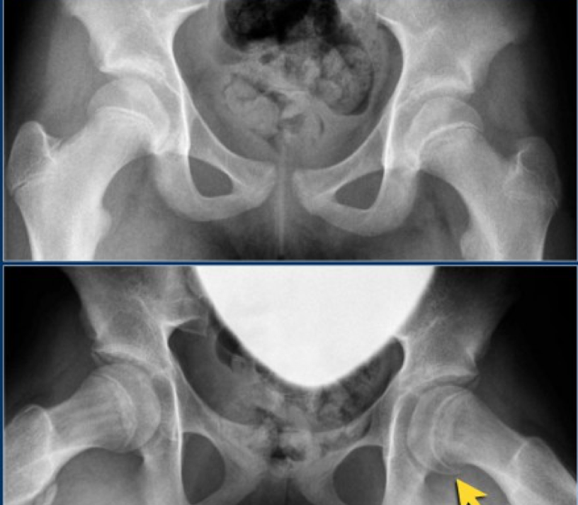
D.     Management      
        i.         Non weight bearing (important to prevent further slippage)    
        ii.         Operative management with possible prophylactic pinning of contralateral hip
E.     Complications      
        i.         Avascular necrosis    
        ii.         Premature closure of physis     
        iii.         Limited ROM   
        iv.         Osteoarthritis

​5.   Legg-calve-perthes disease
A.     Idiopathic avascular necrosis of proximal femoral epiphysis
B.     Usually unilateral (10-15% bilateral)
C.     Clinical features      
        i.         Insidious onset of mild hip or knee pain, possible limp    
        ii.         Repeated episodes of ischemia of femoral head à infarction and necrosis à subchondral stress fracture
D.    Diagnosis      
        i.         Physical exam
                  1.     Decreased abduction and internal rotation
                  2.     Possible thigh atrophy on affected side
                  3.     Limb shortening (advanced cases)    
       ii.         Imaging
                  1.     Early radiographs may be normal, but MRI will show edema in femoral epiphysis with T1 weighted 
Picture
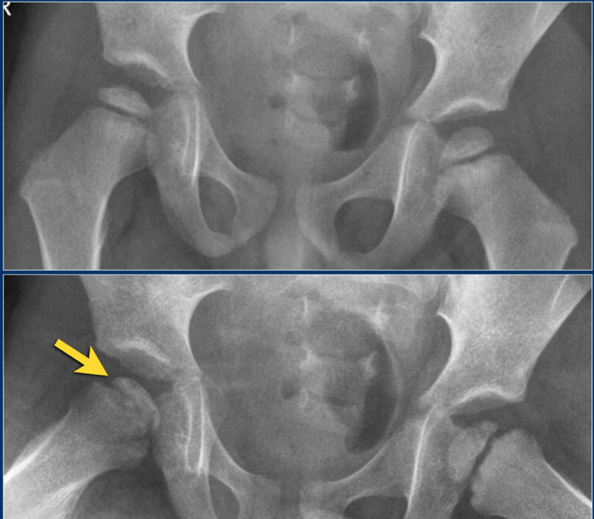
                  2. Radiographic progression of disease
Picture
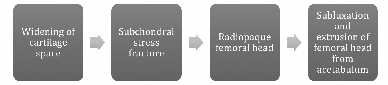
E.     Management      
        i.         Symptomatic tx    
        ii.         Non weight bearing   
        iii.        Operative management may prevent early osteoarthritis
                    can be managed non-operatively with spontaneous revascularization of femoral head
F.     Complications      
        i.         Osteoarthritis    
​        ii.         Coxa magna deformity (short, broad femoral head and column)
Picture

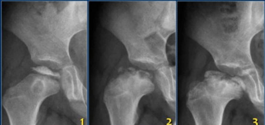
Flynn, John M, and Roger F Widmann. “The Limping Child: Evaluation and Diagnosis.” Journal of the American Academy of Orthopaedic Surgeons, vol. 9, no. 2, Apr. 2001.Horowitz, Diane Lewis, et al. “Approach to Septic Arthritis.” American Family Physician, 15 Sept. 2011, www.aafp.org/afp/2011/0915/p653.html.“Imaging in Slipped Capital Femoral Epiphysis .” Imaging in Slipped Capital Femoral Epiphysis: Practice Essentials, Radiography, Computed Tomography, 6 May 2017, emedicine.medscape.com/article/413810-overview.“Legg-Calve-Perthes.” Legg-Calve-Perthes - Orthopedics - Medbullets Step 2/3, step2.medbullets.com/orthopedics/120519/legg-calve-perthes.Radiology In Ped Emerg Med, Vol 4, Case 17, www.hawaii.edu/medicine/pediatrics/pemxray/v4c17.htmlSwaminathan, Anand. “Pediatric Septic Hip.” Core EM, coreem.net/core/pediatric-septic-hip/.Tintinalli JE, Stapczynski J, Ma O, Cline DM, Cydulka RK, Meckler GD,  T. Tintinalli's Emergency Medicine: A Comprehensive Study Guide, 7e; 2011 Available at:http://accessmedicine.mhmedical.com/content.aspx?bookid=348&sectionid=40381606 Accessed: February 12, 2018
0 Comments

    Orthopedics Blog

    Picture

    Author 

    CMC ER Residents 
    (for learning purposes, imagine all cases present to a free standing ED for dispo and treatment options)

    Subscribe To Blog

    Archives

    June 2018
    April 2018
    March 2018
    October 2017
    September 2017
    February 2016
    January 2016
    December 2015
    November 2015
    October 2015
    September 2015
    August 2015
    July 2015
    June 2015
    May 2015
    April 2015
    March 2015
    February 2015
    January 2015
    December 2014
    November 2014

    Categories

    All
    Lower Extremity
    Nerve Injury
    Spine
    Sports Medicine
    Upper Extremity
    Vascular Injury

    RSS Feed

    Disclaimer: All images and x-rays included on this blog are the sole property of CMC EM Residency and cannot be used or reproduced without written permission.  Patient identifiers have been redacted/changed or patient consent has been obtained.  Information contained in this blog is the opinion of the author and application of material contained in this blog is at the discretion of the practitioner to verify for accuracy.
Powered by Create your own unique website with customizable templates.
  • RESIDENCY
    • About CMC
    • Curriculum
    • Benefits
    • Explore Charlotte
    • Official Site
  • FELLOWSHIP
    • EMS
    • Global EM
    • Pediatric EM
    • Toxicology >
      • Tox Faculty
      • Tox Application
    • (All Others)
  • PEOPLE
    • Program Leadership
    • PGY-3
    • PGY-2
    • PGY-1
    • Alumni
  • STUDENTS/APPLICANTS
    • Medical Students at CMC
    • EM Acting Internship
    • Healthcare Disparities Externship
    • Resident Mentorship
  • #FOAMed
    • EM GuideWire
    • CMC Imaging Mastery
    • Pediatric EM Morsels
    • Blogs, etc. >
      • CMC ECG Masters
      • Core Concepts
      • Cardiology Blog
      • Dr. Patel's Coding Blog
      • Global Health Blog
      • Ortho Blog
      • Pediatric Emergency Medicine
      • Tox Blog
  • Chiefs Corner
    • Top 20
    • Current Chiefs
    • Schedules >
      • Conference/Flashpoint
      • Block Schedule
      • ED Shift Schedule
      • AEC Moonlighting
      • Journal Club/OBP/Audits Schedule
      • Simulation
    • Resources >
      • Fox Reference Library
      • FlashPoint
      • Airway Lecture
      • Student Resources
      • PGY - 1
      • PGY - 2
      • PGY - 3
      • Simulation Reading
      • Resident Wellness
      • Resident Research
      • Resume Builder
    • Individualized Interactive Instruction