CMC COMPENDIUM
  • RESIDENCY
    • About CMC
    • Curriculum
    • Benefits
    • Explore Charlotte
    • Official Site
  • FELLOWSHIP
    • EMS
    • Global EM
    • Pediatric EM
    • Toxicology >
      • Tox Faculty
      • Tox Application
    • (All Others)
  • PEOPLE
    • Program Leadership
    • PGY-3
    • PGY-2
    • PGY-1
    • Alumni
  • STUDENTS/APPLICANTS
    • Medical Students at CMC
    • EM Acting Internship
    • Healthcare Disparities Externship
    • Resident Mentorship
  • #FOAMed
    • EM GuideWire
    • CMC Imaging Mastery
    • Pediatric EM Morsels
    • Blogs, etc. >
      • CMC ECG Masters
      • Core Concepts
      • Cardiology Blog
      • Dr. Patel's Coding Blog
      • Global Health Blog
      • Ortho Blog
      • Pediatric Emergency Medicine
      • Tox Blog
  • Chiefs Corner
    • Top 20
    • Current Chiefs
    • Schedules >
      • Conference/Flashpoint
      • Block Schedule
      • ED Shift Schedule
      • AEC Moonlighting
      • Journal Club/OBP/Audits Schedule
      • Simulation
    • Resources >
      • Fox Reference Library
      • FlashPoint
      • Airway Lecture
      • Student Resources
      • PGY - 1
      • PGY - 2
      • PGY - 3
      • Simulation Reading
      • Resident Wellness
      • Resident Research
      • Resume Builder
    • Individualized Interactive Instruction

Pediatric Elbow Dislocation

1/23/2015

0 Comments

 
HPI:  Pediatric male presents after a fall onto outstretched arm with obvious deformity to right elbow.  

PE:  Patient is unable to cross fingers and has paresthesias in the ulnar nerve distribution.  Initial radiograph showed posterior dislocation with questionable hyperdensity representing the medial epicondyle.  We were unable to reduce under fluoroscopy as the medial epicondyle was more visible and the patient eventually required ORIF.

Imaging: AP and lateral radiograph of the elbow

Picture
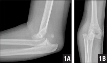
Image 1: Showing associated medial epicondyle fracture
How to Diagnosis: Two important lines help in making the diagnosis; radiocapitella and anterior humeral.

Radiocapitellar line:  Drawn through the center of the radial neck and should pass the center of the capitellum as shown below
Picture
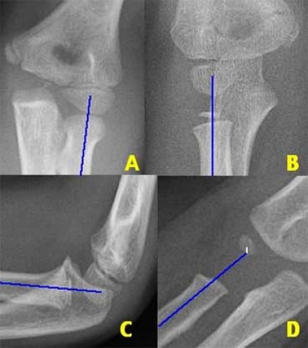
Anterior humeral line:  Drawn through the lateral view of the surface of the humerus and should pass through the middle of the capitellum
Picture
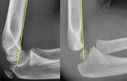
Ossification: 6 sites that fuse at different ages.  Mnemonic = CRITOE
  • C - Capitellum
  • R - Radius
  • I - Internal (medial) epicondyle
  • T - Trochlea
  • O - Olecranon
  • E - External (lateral) epicondyle
Ossify around 1-3-4-7-9-11
 
Treatment: Nonoperative with closed reduction and early range of motion (1-2 weeks) is most common.  ORIF required for incarcerated medial epicondyle, inability to obtain close reduction, or significant instability.

Pearls:
  • Most common mechanism is fall on outstretched hand
  • Reduction technique involves gradual axial traction starting in full extension and moving towards full flexion
  • Consider child abuse in all cases if story does not fit injury
  • Brachial artery and median nerve most commonly injured
  • Ulnar nerve injury more common with medial epicondyle avulsion

0 Comments

Your comment will be posted after it is approved.


Leave a Reply.

    Orthopedics Blog

    Picture

    Author 

    CMC ER Residents 
    (for learning purposes, imagine all cases present to a free standing ED for dispo and treatment options)

    Subscribe To Blog

    Archives

    June 2018
    April 2018
    March 2018
    October 2017
    September 2017
    February 2016
    January 2016
    December 2015
    November 2015
    October 2015
    September 2015
    August 2015
    July 2015
    June 2015
    May 2015
    April 2015
    March 2015
    February 2015
    January 2015
    December 2014
    November 2014

    Categories

    All
    Lower Extremity
    Nerve Injury
    Spine
    Sports Medicine
    Upper Extremity
    Vascular Injury

    RSS Feed

    Disclaimer: All images and x-rays included on this blog are the sole property of CMC EM Residency and cannot be used or reproduced without written permission.  Patient identifiers have been redacted/changed or patient consent has been obtained.  Information contained in this blog is the opinion of the author and application of material contained in this blog is at the discretion of the practitioner to verify for accuracy.
Powered by Create your own unique website with customizable templates.
  • RESIDENCY
    • About CMC
    • Curriculum
    • Benefits
    • Explore Charlotte
    • Official Site
  • FELLOWSHIP
    • EMS
    • Global EM
    • Pediatric EM
    • Toxicology >
      • Tox Faculty
      • Tox Application
    • (All Others)
  • PEOPLE
    • Program Leadership
    • PGY-3
    • PGY-2
    • PGY-1
    • Alumni
  • STUDENTS/APPLICANTS
    • Medical Students at CMC
    • EM Acting Internship
    • Healthcare Disparities Externship
    • Resident Mentorship
  • #FOAMed
    • EM GuideWire
    • CMC Imaging Mastery
    • Pediatric EM Morsels
    • Blogs, etc. >
      • CMC ECG Masters
      • Core Concepts
      • Cardiology Blog
      • Dr. Patel's Coding Blog
      • Global Health Blog
      • Ortho Blog
      • Pediatric Emergency Medicine
      • Tox Blog
  • Chiefs Corner
    • Top 20
    • Current Chiefs
    • Schedules >
      • Conference/Flashpoint
      • Block Schedule
      • ED Shift Schedule
      • AEC Moonlighting
      • Journal Club/OBP/Audits Schedule
      • Simulation
    • Resources >
      • Fox Reference Library
      • FlashPoint
      • Airway Lecture
      • Student Resources
      • PGY - 1
      • PGY - 2
      • PGY - 3
      • Simulation Reading
      • Resident Wellness
      • Resident Research
      • Resume Builder
    • Individualized Interactive Instruction