CMC COMPENDIUM
  • RESIDENCY
    • About CMC
    • Curriculum
    • Benefits
    • Explore Charlotte
    • Official Site
  • FELLOWSHIP
    • EMS
    • Global EM
    • Pediatric EM
    • Toxicology >
      • Tox Faculty
      • Tox Application
    • (All Others)
  • PEOPLE
    • Program Leadership
    • PGY-3
    • PGY-2
    • PGY-1
    • Alumni
  • STUDENTS/APPLICANTS
    • Medical Students at CMC
    • EM Acting Internship
    • Healthcare Disparities Externship
    • Resident Mentorship
  • #FOAMed
    • EM GuideWire
    • CMC Imaging Mastery
    • Pediatric EM Morsels
    • Blogs, etc. >
      • CMC ECG Masters
      • Core Concepts
      • Cardiology Blog
      • Dr. Patel's Coding Blog
      • Global Health Blog
      • Ortho Blog
      • Pediatric Emergency Medicine
      • Tox Blog
  • Chiefs Corner
    • Top 20
    • Current Chiefs
    • Schedules >
      • Conference/Flashpoint
      • Block Schedule
      • ED Shift Schedule
      • AEC Moonlighting
      • Journal Club/OBP/Audits Schedule
      • Simulation
    • Resources >
      • Fox Reference Library
      • FlashPoint
      • Airway Lecture
      • Student Resources
      • PGY - 1
      • PGY - 2
      • PGY - 3
      • Simulation Reading
      • Resident Wellness
      • Resident Research
      • Resume Builder
    • Individualized Interactive Instruction

Patella  Fractures

5/18/2015

0 Comments

 
HPI:
20ish year old and otherwise healthy male presents as the restrained driver in head-on MVC at low speed just prior to arrival. He has a large linear laceration to anterior aspect of R knee and is unable to fully extend the knee. Wound is hemostatic. No other injuries or complaints.

Physical Examination:
10cm linear, horizontal laceration to the anterior aspect of the patient’s R knee.  Knee is held in slightly flexed positioning. Decreased ROM of the knee secondary to pain and injury, patient unable to actively extend extremity in straight leg raise.  FROM of ankle. 2+ DP and PT pulses present. Sensation intact to light touch throughout. No erythema, swelling or significant tenderness present over RLE. 

Radiology:

AP/Lateral/Sunrise/Oblique views of R knee:
Transverse fracture of the patella with 3mm of displacement present and surrounding soft tissue edema

Management:
  • Bedside washout of wound with copious saline and with wet gauze placement
  • Tetanus updated
  • 2g IV Ancef administered
  • Admission for ORIF of R patella with tension band construction

Discussion:
  • Patella = largest sesamoid bone in the body with the thickest articular cartilage 
  • Patella fractures = 1% of all bony injuries
  • Injury usually by direct impact or indirect eccentric contraction of quadriceps with hyperextension of the knee
  • Usually seen in younger adult population (20-50 years old)

 Types of fracture:
Picture
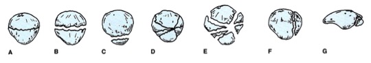
Figure obtained from jaaos.org; Patellar Fractures in Adults; AAOS April 2011 vol. 19 no. 4 198-207,Figure 4
A)  Nondisplaced
B)  Transverse
C)  Pole or sleeve (upper or lower) 
D)  Comminuted nondisplaced
E)   Comminuted displaced
F)   Vertical
G)  Osteochondral

Evaluate with physical examination first
Severe knee pain and soft tissue swelling should raise suspicion for injury
Concerning findings:
  • Palpable bony defect
  • Large hemarthrosis
  • High riding patella
  • Inability to weight bear
  • Active extension of knee difficult to perform
  • Unable to perform straight leg raise test = retinaculum disrupted (the connective tissue band around tendons that hold them in place)
Picture
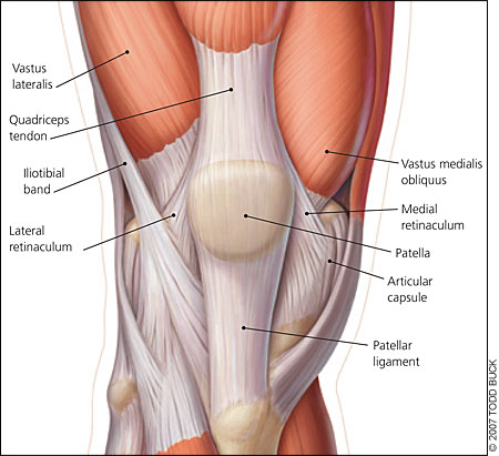
Image obtained from aafp.org, Am Fam Physician. 2007 Jan 15;75(2):194-202.
Obtain Radiographs
Fracture displacement best viewed on lateral films, but obtain AP/Lat/Oblique/Sunrise to fully evaluate
Degree of displacement =  degree of retinacular disruption

Treatment:
Nonoperative:
  • If intact extensor mechanism
  • Nondisplaced or minimally displaced fracture
  • Vertical fracture
Knee immobilized in extension with brace or cylinder cast with early WBAT
Early active ROM with hinged knee brace after 2-3 weeks

Operative:
  • Open fractures
  • Extensor mechanism failure
  • Fracture with articular displacement of >2mm
  • Displaced patella fracture >3mm
  • Patella sleeve fractures in children
Usually managed by ORIF with tension band construction

Pearls:
Watch out for patella sleeve fractures in peds – need high index of suspicion to diagnose
  • Small avulsion fractures that occur between cartilage “sleeve” and main bony patella usually in kids 8-12 yrs old when patellar ossification is nearly complete
Consider obtaining MRI in a child with normal x-rays but who is unable to straight leg raise

Bipartite patella = failure of bony fusion
  • Anatomic anomaly seen in up to 8% of population and may be mistaken for fracture
  • May be bilateral in up to 50% of people with this variation

Any lacerations present must be assumed to communicate with the knee joint until this assumption is disproved by a saline load joint challenge

A patellar fracture is problematic and requires intervention if extensor mechanism of knee is nonfunctional = unable to perform straight leg raise test
0 Comments

Your comment will be posted after it is approved.


Leave a Reply.

    Orthopedics Blog

    Picture

    Author 

    CMC ER Residents 
    (for learning purposes, imagine all cases present to a free standing ED for dispo and treatment options)

    Subscribe To Blog

    Archives

    June 2018
    April 2018
    March 2018
    October 2017
    September 2017
    February 2016
    January 2016
    December 2015
    November 2015
    October 2015
    September 2015
    August 2015
    July 2015
    June 2015
    May 2015
    April 2015
    March 2015
    February 2015
    January 2015
    December 2014
    November 2014

    Categories

    All
    Lower Extremity
    Nerve Injury
    Spine
    Sports Medicine
    Upper Extremity
    Vascular Injury

    RSS Feed

    Disclaimer: All images and x-rays included on this blog are the sole property of CMC EM Residency and cannot be used or reproduced without written permission.  Patient identifiers have been redacted/changed or patient consent has been obtained.  Information contained in this blog is the opinion of the author and application of material contained in this blog is at the discretion of the practitioner to verify for accuracy.
Powered by Create your own unique website with customizable templates.
  • RESIDENCY
    • About CMC
    • Curriculum
    • Benefits
    • Explore Charlotte
    • Official Site
  • FELLOWSHIP
    • EMS
    • Global EM
    • Pediatric EM
    • Toxicology >
      • Tox Faculty
      • Tox Application
    • (All Others)
  • PEOPLE
    • Program Leadership
    • PGY-3
    • PGY-2
    • PGY-1
    • Alumni
  • STUDENTS/APPLICANTS
    • Medical Students at CMC
    • EM Acting Internship
    • Healthcare Disparities Externship
    • Resident Mentorship
  • #FOAMed
    • EM GuideWire
    • CMC Imaging Mastery
    • Pediatric EM Morsels
    • Blogs, etc. >
      • CMC ECG Masters
      • Core Concepts
      • Cardiology Blog
      • Dr. Patel's Coding Blog
      • Global Health Blog
      • Ortho Blog
      • Pediatric Emergency Medicine
      • Tox Blog
  • Chiefs Corner
    • Top 20
    • Current Chiefs
    • Schedules >
      • Conference/Flashpoint
      • Block Schedule
      • ED Shift Schedule
      • AEC Moonlighting
      • Journal Club/OBP/Audits Schedule
      • Simulation
    • Resources >
      • Fox Reference Library
      • FlashPoint
      • Airway Lecture
      • Student Resources
      • PGY - 1
      • PGY - 2
      • PGY - 3
      • Simulation Reading
      • Resident Wellness
      • Resident Research
      • Resume Builder
    • Individualized Interactive Instruction