CMC COMPENDIUM
  • Prospective Applicants
    • 2020 MATCH
    • About CMC
    • Our Curriculum
    • Our Residents
    • Our Fellowships
    • Program Leadership
    • Explore Charlotte
    • Official Site
  • ROTATING STUDENTS
    • Prospective Visiting Students
    • UNC Students
    • Healthcare Disparities Externship
    • Current Students
  • Current Residents
    • Airway Lecture!
    • PGY - 1
    • PGY - 2
    • PGY - 3
    • Simulation Reading
    • Blogs >
      • EM GuideWire
      • CMC ECG Masters
      • Core Concepts
      • #FOAMed
      • Cardiology Blog
      • Dr. Patel's Coding Blog
      • Global Health Blog
      • Ortho Blog
      • Pediatric Emergency Medicine
      • Tox Blog
    • Board Review
    • Journal Club
    • Resident Wellness
    • Resident Research
  • Top 20
  • Chiefs Corner
    • Schedules >
      • Conference/Flashpoint
      • Block Schedule
      • ED Shift Schedule
      • AEC Moonlighting
      • Journal Club/OBP/Audits Schedule
      • Simulation
    • Individualized Interactive Instruction
    • Evaluations/Interview Season
    • Contact Info
    • Resume Builder

The Wrist

3/5/2018

0 Comments

 
Authored by Dr. Javier Andrade
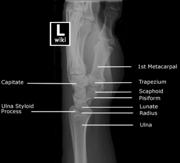
  • With any orthopedic injury, it is important to understand the normal bony anatomy of the injured area you are examining:
    • The Wrist is a complex area with multiples bones and joint.
    • It is composed of the more proximal ulna and radius which anchor the wrist and help create the supination and pronation needed for complex hand movements
    • The distal portion of the wrist is made up of the 8 carpal bones arranged in two rows.
Picture
Picture
  • Now that we now what bones make up the wrist, we can talk a bit more about the alignment and tricks we can use to see if there is an injury to the wrist.
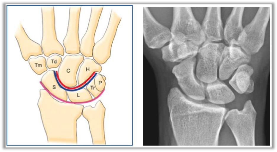
    • One such trick we can use for the PA view are Gilula’s Line. It is a technique which uses 3 curved lines to check the alignment of the carpal bone rows.
      • Proximal row, proximal portion (pink)
      • Proximal row, distal portion (blue)
      • Distal row, proximal portion (red)
Picture
  • Also, as with any ortho injury, remember to assess that the patient is neurovascularly intact!
    • Vascular: Can use the doppler probe to assess
      • Ulnar artery
      • Radial Artery
      • Palmar arch
    • Nerves:
      • Radial n: Test the extensor pollicus longus using “thumbs up”
      • Median n: Test using the “OK sign”
        • Make sure patient flexes at the DIP joint
      • Ulnar n: Test using pinky abduction
  • Pathology:
    • Scaphoid Fx
      • Most commonly injured carpal bone and can be easily missed. If you have clinical exam findings but no radiologic findings, treat this as a fracture!
      • Tx: Thumb spica cast and reimage in 12 days.
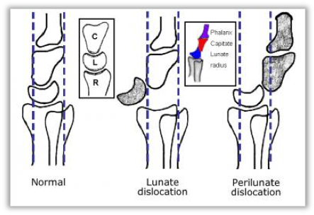
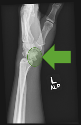
    • Lunate and Perilunate dislocation
      • Associated with high-energy mechanism of injury as it has to tear through multiple ligaments that hold the hand together. Be on the lookout for other injuries in the wrist.
        • Associated with median n injury in 25% of patients
      • Notice the anatomy on the normal image (left). We can use the lines on the lateral XR view to assess for injury
Picture
Picture
  • Hamate fracture:
    • Body: Clenched fist against solid object
      • Typically requires surgical intervention
    • Hook: Associated with injuries that require swinging equipment (baseball, tennis, etc.)
      • Can cause ulnar n. injury or impingement
  • Distal Radius Fractures:
    • Colle’s: Fx with posterior displacement
    • Smith’s: Fx with anterior displacement
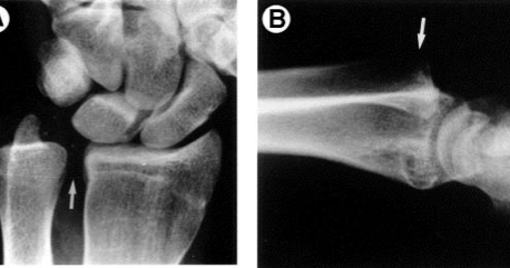
  • Distal Radial-Ulnar joint (DRUJ) injuries
    • Normally, the ulna sits on a small groove on the distal radius known as the sigmoid notch.
      • Any spacing here is abnormal
Picture
  • Management:
    • If purely ligamentous closed immobilization and outpatient follow-up
    • If associated with fracture then operative repair is necessary
0 Comments

Your comment will be posted after it is approved.


Leave a Reply.

    Orthopedics Blog

    Picture

    Author 

    CMC ER Residents 
    (for learning purposes, imagine all cases present to a free standing ED for dispo and treatment options)

    Subscribe To Blog

    Archives

    June 2018
    April 2018
    March 2018
    October 2017
    September 2017
    February 2016
    January 2016
    December 2015
    November 2015
    October 2015
    September 2015
    August 2015
    July 2015
    June 2015
    May 2015
    April 2015
    March 2015
    February 2015
    January 2015
    December 2014
    November 2014

    Categories

    All
    Lower Extremity
    Nerve Injury
    Spine
    Sports Medicine
    Upper Extremity
    Vascular Injury

    RSS Feed

    Disclaimer: All images and x-rays included on this blog are the sole property of CMC EM Residency and cannot be used or reproduced without written permission.  Patient identifiers have been redacted/changed or patient consent has been obtained.  Information contained in this blog is the opinion of the author and application of material contained in this blog is at the discretion of the practitioner to verify for accuracy.
Disclaimer: All original material and images included on this website are the sole property of CMC EM Residency and cannot be used or reproduced without written permission.  Information contained on this website is the opinion of the authors and does not necessarily represent the official opinion of Atrium Health or Carolinas Emergency Medicine Residency. 


For Health Care Providers:  Every effort is made to provide the most up to date evidence based medicine.  However, this content may not necessarily reflect the standard of care and application of material contained on this website is at the discretion of the practitioner to verify for accuracy.


For the Public:  This content is not intended to be a substitute for professional medical advice, diagnosis, or treatment.  Relying on information contained on this website is done at your own risk.  Do not disregard professional medical advice or delay seeking care secondary to content on this website.  Call 911 or seek a medical professional immediately for any medical emergencies.
Like us on Facebook or
follow us on Twitter/Instagram

Contact Us:

Department of Emergency Medicine
Medical Education Building., Third floor
1000 Blythe Blvd.
Charlotte, NC 28203

Telephone: 704-355-3658 
Fax: 704-355-7047
​
  • Prospective Applicants
    • 2020 MATCH
    • About CMC
    • Our Curriculum
    • Our Residents
    • Our Fellowships
    • Program Leadership
    • Explore Charlotte
    • Official Site
  • ROTATING STUDENTS
    • Prospective Visiting Students
    • UNC Students
    • Healthcare Disparities Externship
    • Current Students
  • Current Residents
    • Airway Lecture!
    • PGY - 1
    • PGY - 2
    • PGY - 3
    • Simulation Reading
    • Blogs >
      • EM GuideWire
      • CMC ECG Masters
      • Core Concepts
      • #FOAMed
      • Cardiology Blog
      • Dr. Patel's Coding Blog
      • Global Health Blog
      • Ortho Blog
      • Pediatric Emergency Medicine
      • Tox Blog
    • Board Review
    • Journal Club
    • Resident Wellness
    • Resident Research
  • Top 20
  • Chiefs Corner
    • Schedules >
      • Conference/Flashpoint
      • Block Schedule
      • ED Shift Schedule
      • AEC Moonlighting
      • Journal Club/OBP/Audits Schedule
      • Simulation
    • Individualized Interactive Instruction
    • Evaluations/Interview Season
    • Contact Info
    • Resume Builder