CMC COMPENDIUM
  • RESIDENCY
    • About CMC
    • Curriculum
    • Benefits
    • Explore Charlotte
    • Official Site
  • FELLOWSHIP
    • EMS
    • Global EM
    • Pediatric EM
    • Toxicology >
      • Tox Faculty
      • Tox Application
    • (All Others)
  • PEOPLE
    • Program Leadership
    • PGY-3
    • PGY-2
    • PGY-1
    • Alumni
  • STUDENTS/APPLICANTS
    • Medical Students at CMC
    • EM Acting Internship
    • Healthcare Disparities Externship
    • Resident Mentorship
  • #FOAMed
    • EM GuideWire
    • CMC Imaging Mastery
    • Pediatric EM Morsels
    • Blogs, etc. >
      • CMC ECG Masters
      • Core Concepts
      • Cardiology Blog
      • Dr. Patel's Coding Blog
      • Global Health Blog
      • Ortho Blog
      • Pediatric Emergency Medicine
      • Tox Blog
  • Chiefs Corner
    • Top 20
    • Current Chiefs
    • Schedules >
      • Conference/Flashpoint
      • Block Schedule
      • ED Shift Schedule
      • AEC Moonlighting
      • Journal Club/OBP/Audits Schedule
      • Simulation
    • Resources >
      • Fox Reference Library
      • FlashPoint
      • Airway Lecture
      • Student Resources
      • PGY - 1
      • PGY - 2
      • PGY - 3
      • Simulation Reading
      • Resident Wellness
      • Resident Research
      • Resume Builder
    • Individualized Interactive Instruction

The FOot

6/12/2018

0 Comments


By: DR. AUSTIN COSTA

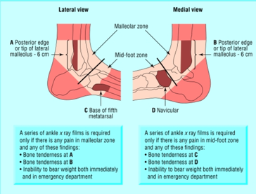
When to order foot X-rays?:
  • Ottawa midfoot rules
    • Pain with palpation of base of 5thmetatarsal
    • Pain with palpation of navicular
    • Inability to bear weight both at time of injury and in ED
  • Suspected injury with high energy mechanism 
Picture
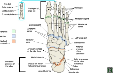
Anatomy (From proximal to distal):
  • Hindfoot 
    • Calcaneus and talus
  • Chopart joint 
  • Midfoot 
    • Navicular, medial cuneiform, middle cuneiform, lateral cuneiform, and cuboid
  • Lizfranc joint
    • Lizfranc ligament connects medial cuneiform and 2ndMT on plantar aspect of foot
  • Forefoot 
    • Metatarsals, phalanges
Picture
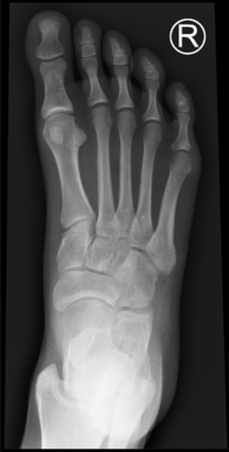
​Standard Views:
  • AP
    • Forefoot 
    • Midfoot
    • Lizfranc injury
      • Alignment of 1stMT and medial cunneaform
      • Alignment of 2ndMT and middle cunneaform
Picture
Oblique
  • Forefoot
  • Midfoot
  • Calcaneus 
  • Alignment for 3rdMT with lateral cuneiform
  • Alignment of 4thMT with cuboid
Picture
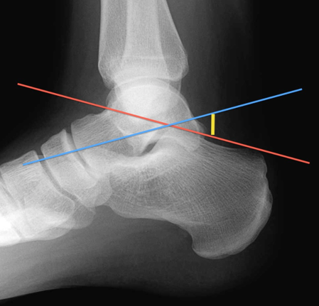
  • Lateral
    • Hindfoot 
      • Boehler’s angle
        • upper edge of the calcaneal body posteriorly to the upper edge of the posterior articular facet of the calcaneus (red)
        • upper edge of the posterior articular facet of the calcaneus to the upper edge of the anterior process of the calcaneus (blue)
        • Normal is 25-40 degress ​
Picture
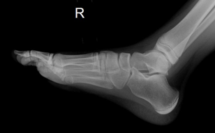
  • Dorsal foot arch
    • Weight bearing films (if able to tolerate)
    • Allows spreading of metatarsals
Picture
  • 1. ​Approach to foot X-rays:
    • Assess cortical disruption for fractures in area of tenderness
      • Forefoot
        • AP, oblique
      • Midfoot
        • AP, oblique
      • Hindfoot
        • Oblique, lateral
        • Boehler’s angle < 25 degress = calcaneal depression fracture
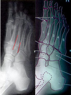
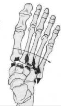
  •  2. Lizfranc Injury
    • Plantar ecchymosis
    • Stress tarso-MT joint
    • Get weight bearing films!
    • Instability ED Ortho consult
Picture
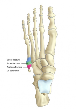
  • 3. Base of 5thMetatarsal 
    • Pseudo-Jone(Zones)
      • Ankle inversion
      • Peroneus brevis insertion
    • Jones (Zone II) 
      • Metaphyseal-diaphyseal junction
      • At level of 4th-5thmetatarsal joint
      • High risk of non-union
    • Diaphyseal Stress Fracture (Zone III)
      • Repetitive microtrauma
Picture
Management:
  • Hindfoot: Splint (Bulky Jones), NWB
    • Boehler’s angle
    • Look for spinal injuries
  • Midfoot: Splint (sugar tong w/ posterior slab), NWB
    • Lizfranc. -> Weight bearing films
  • Forefoot: 
    • Metatarsals: CAM boot, NWB -> WBAT. Except 1stand base of 5th zones 2&3
    • Phylanges: Buddy tape, post op shoe, unless significantly displaced/rotated
References
  1. Londner M, Hammer D, Kelen GD. Foot injuries. In: Tintinalli JE, Kelen GD, Stapczynski JS, Ma, OJ, Cline DM, editors. Tintinalli’s Emergency Medicine. 8 th ed. New York: McGraw-Hill; 2004; (cited 2017, Sep 08)
  2. Smith, M. S., Hatch, R., & Eiff, M. (2012). Calcaneous and Other Tarsal Fractures; Metatarsal Fractures; Toe Fractures; . In Fracture Management for Primary Care (3rd ed., pp. 276-326). Philadelphia, PA: Saunders.
  3. Weatherford, B. (n.d.). 5th Metatarsal Base Fracture. Retrieved September 08, 2017, from http://www.orthobullets.com/foot-and-ankle/7031/5th-metatarsal-base-fracture
  4. Hatch, R. L., & Hacking, S. (2003, December 15). Evaluation and Management of Toe Fractures. Retrieved September 08, 2017, from http://www.aafp.org/afp/2003/1215/p2413.html
  5. Foot Fracture Treatment & Management. (2017, August 03). Retrieved September 08, 2017, from http://emedicine.medscape.com/article/825060-treatment#d10
0 Comments

Your comment will be posted after it is approved.


Leave a Reply.

    Orthopedics Blog

    Picture

    Author 

    CMC ER Residents 
    (for learning purposes, imagine all cases present to a free standing ED for dispo and treatment options)

    Subscribe To Blog

    Archives

    June 2018
    April 2018
    March 2018
    October 2017
    September 2017
    February 2016
    January 2016
    December 2015
    November 2015
    October 2015
    September 2015
    August 2015
    July 2015
    June 2015
    May 2015
    April 2015
    March 2015
    February 2015
    January 2015
    December 2014
    November 2014

    Categories

    All
    Lower Extremity
    Nerve Injury
    Spine
    Sports Medicine
    Upper Extremity
    Vascular Injury

    RSS Feed

    Disclaimer: All images and x-rays included on this blog are the sole property of CMC EM Residency and cannot be used or reproduced without written permission.  Patient identifiers have been redacted/changed or patient consent has been obtained.  Information contained in this blog is the opinion of the author and application of material contained in this blog is at the discretion of the practitioner to verify for accuracy.
Powered by Create your own unique website with customizable templates.
  • RESIDENCY
    • About CMC
    • Curriculum
    • Benefits
    • Explore Charlotte
    • Official Site
  • FELLOWSHIP
    • EMS
    • Global EM
    • Pediatric EM
    • Toxicology >
      • Tox Faculty
      • Tox Application
    • (All Others)
  • PEOPLE
    • Program Leadership
    • PGY-3
    • PGY-2
    • PGY-1
    • Alumni
  • STUDENTS/APPLICANTS
    • Medical Students at CMC
    • EM Acting Internship
    • Healthcare Disparities Externship
    • Resident Mentorship
  • #FOAMed
    • EM GuideWire
    • CMC Imaging Mastery
    • Pediatric EM Morsels
    • Blogs, etc. >
      • CMC ECG Masters
      • Core Concepts
      • Cardiology Blog
      • Dr. Patel's Coding Blog
      • Global Health Blog
      • Ortho Blog
      • Pediatric Emergency Medicine
      • Tox Blog
  • Chiefs Corner
    • Top 20
    • Current Chiefs
    • Schedules >
      • Conference/Flashpoint
      • Block Schedule
      • ED Shift Schedule
      • AEC Moonlighting
      • Journal Club/OBP/Audits Schedule
      • Simulation
    • Resources >
      • Fox Reference Library
      • FlashPoint
      • Airway Lecture
      • Student Resources
      • PGY - 1
      • PGY - 2
      • PGY - 3
      • Simulation Reading
      • Resident Wellness
      • Resident Research
      • Resume Builder
    • Individualized Interactive Instruction