CMC COMPENDIUM
  • RESIDENCY
    • About CMC
    • Curriculum
    • Benefits
    • Explore Charlotte
    • Official Site
  • FELLOWSHIP
    • EMS
    • Global EM
    • Pediatric EM
    • Toxicology >
      • Tox Faculty
      • Tox Application
    • (All Others)
  • PEOPLE
    • Program Leadership
    • PGY-3
    • PGY-2
    • PGY-1
    • Alumni
  • STUDENTS/APPLICANTS
    • Medical Students at CMC
    • EM Acting Internship
    • Healthcare Disparities Externship
    • Resident Mentorship
  • #FOAMed
    • EM GuideWire
    • CMC Imaging Mastery
    • Pediatric EM Morsels
    • Blogs, etc. >
      • CMC ECG Masters
      • Core Concepts
      • Cardiology Blog
      • Dr. Patel's Coding Blog
      • Global Health Blog
      • Ortho Blog
      • Pediatric Emergency Medicine
      • Tox Blog
  • Chiefs Corner
    • Top 20
    • Current Chiefs
    • Schedules >
      • Conference/Flashpoint
      • Block Schedule
      • ED Shift Schedule
      • AEC Moonlighting
      • Journal Club/OBP/Audits Schedule
      • Simulation
    • Resources >
      • Fox Reference Library
      • FlashPoint
      • Airway Lecture
      • Student Resources
      • PGY - 1
      • PGY - 2
      • PGY - 3
      • Simulation Reading
      • Resident Wellness
      • Resident Research
      • Resume Builder
    • Individualized Interactive Instruction

The Adult Ankle

10/15/2017

2 Comments

 
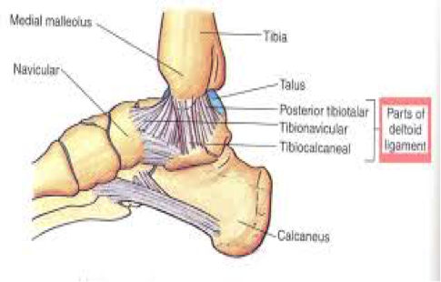
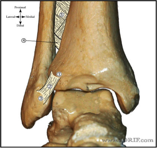
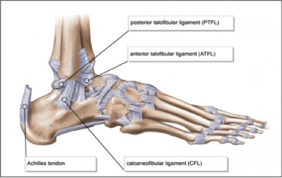
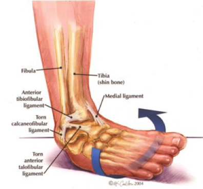
​In order to begin an appropriate assessment of the ankle, it is necessary to review the significant anatomical structures.
 
Bony Anatomy
 
First, we have the two long bones that form the proximal portion of the joint – the tibia and the fibula. At the ankle joint, the tibia extends as the lateral malleolus and posterior malleolus while the fibula forms the medial malleolus. The distal boney structure of the joint is formed by the talus.
Picture
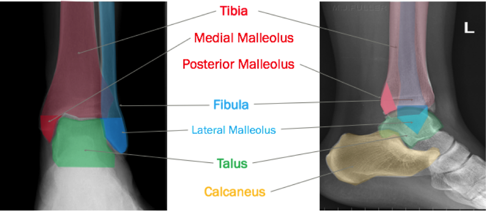
Joint Anatomy
A majority of the articular surface is formed by the horizontal portion of the distal tibia (the tibial plafond) which extends parallel to the dome of the talus; taken with the medial and lateral malleoli, it forms a rectangular socket known as the ankle mortise. Being a synovial joint, the ankle joint (between the ankle mortise and talar dome) is surrounded by a joint capsule. Like the knee joint capsule, the ankle capsule has an additional cranial extension at the syndesmosis.
Picture
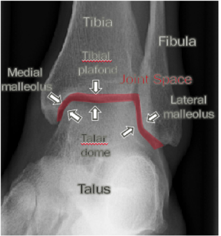
Ligament Anatomy
The lateral and medial malleoli have ligamentous attachments to the talus and calcaneus that contribute to stability of the joint.
  • The lateral ligamentous complex is composed of the posterior talofibular ligament (PTFL), anterior talofibular ligament (ATFL) and the calcaneofibular ligament (CFL). The ATFL is the first or only ligament to be injured in the majority of ankle sprains. Specifically, injuries that result from inversion mechanisms. 
Picture
Picture
  • The medial malleolus is anchored by 3 ligaments collectively referred to as the deltoid ligament. Injury to the deltoid ligament is much less common and disruption of this ligamentous complex conveys a high mechanism of injury. 
Picture
  • Cranially the tibia and fibula are anchored to each other by the fibrous joint of the syndesmosis (along the interosseous membrane).
Picture
Initial Evaluation
A patient presenting with a suspected ankle injury should undergo the following assessment:
  1. Inspection – the joint should be inspected for any signs of ecchymosis, swelling, or skin tenting that could represent injury. Also, be sure to remove any dressing or bandages – you don’t want to miss an open fracture
  2. Palpate – Palpate the entire length of the fibula because syndesmotic injuries can be associated with proximal fibula fracture. Palpate the distal tibia, the bony portions of the foot, and the Achilles tendon. If tenderness of the Achilles is present, squeeze the calf muscles to perform a Thompson test.
  3. Range of Motion -  Check for pain on gentle passive inversion and eversion of the ankle that can indicate a ligament injury. Assess the patient’s ability to plantar and dorsiflex
  4. Deficits – Use 2-point discrimination to test for nerve deficits. An important testing location is between the webbing of the first two digits to assess the deep peroneal nerve. Assess for pulse deficits, make use of the Doppler if you are having difficulty with palpation.   
  5. Joints above and below – Perform an assessment on the joints above and below to detect for any additional injuries.
 
Our initial evaluation is important because it helps utilize the following decision tool:
 
Ottawa Ankle Rule
The Ottawa Ankle Rule helps us screen for patients that will benefit from ankle imaging. Ankle Imaging is warranted in those who have ankle pain and:
  • tenderness over the posterior 6 cm or tip of the posterior or lateral malleolus -  ankle-ray is indicated.
  • patient is unable to take four steps both immediately and in the emergency department -  x-ray of the painful area is indicated
Sensitivities for the Ottawa ankle rule range from the high 90%-100% range for “clinically significant” ankle and midfoot fractures. This is defined as a fracture or an avulsion greater than 3 mm. Specificities for the Ottawa ankle rules are approximately 41% so while it is a good screening tool, we cannot use it to rule in pathology.
 
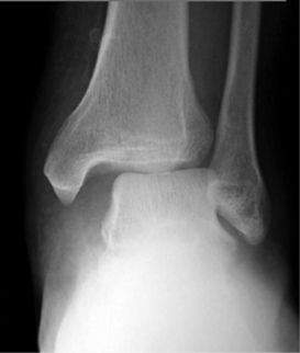
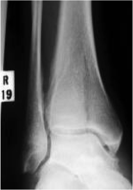
Tips from the creators at University of Ottawa:
Palpate the entire distal 6cm of the fibula and tibia;
Do not neglect the importance of medial malleolar tenderness;
“Bearing weight” counts even if the patient limps;
Be caution in patients under age 18.
 
Imaging Studies
When imaging the ankle obtain 3 views to appropriately evaluate the joint: AP, Lateral and the Mortise study. 
Picture
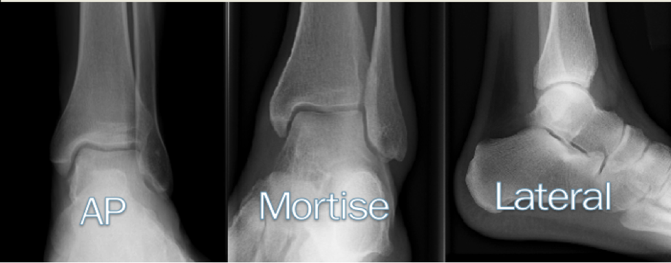
Radiographic Evaluation
Begin with the AP and Lateral views. Trace the entire length of the tibia and fibula paying special attention to the fibula on the lateral view, for oblique fractures may be difficult to see.
 
Next, proceed to the mortise view. Trace around the mortise and Talar dome, evaluate for joint space uniformity. The ring structure of the ankle is made up of three bones (tibia, fibula and talus) and three ligaments (medial and lateral collateral ligaments and interosseous ligament) if there is one break in the ring, look for a second.
 
To evaluate the integrity of the syndesmosis use the following measurements:
•Tibiofibular overlap < 1mm
•Increased medial clear space: less than or equal to 4 mm
•Increased Tibiofibular clear space: < 6 mm
Picture
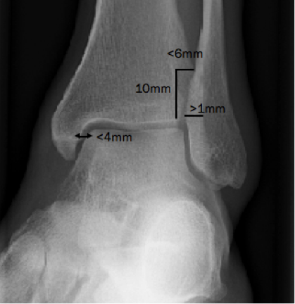
​Additional radiographic measurements
Talo-crural angle : The angle is formed by drawing a line parallel to the tibial plafond, a line perpendicular to the tibail plafond, and a line connecting the lateral and medial malleoli. This angle should be between 75 – 87 degrees and should be within 2-5 degrees of contralateral side. Deviation from these measurements is indicative of fibula shortening.
 
Picture
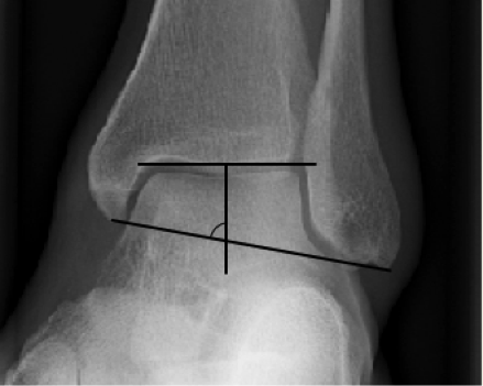
​Typical Fracture Patterns
Isolated Medial Malleolus Fracture  
Picture
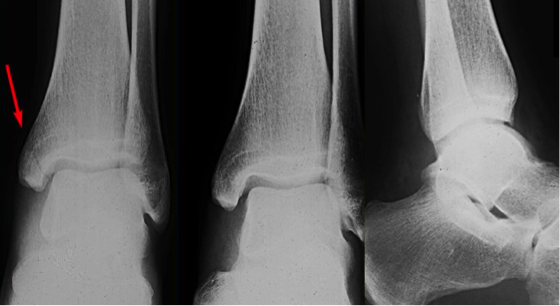
Isolated Lateral Malleolus Fracture
Picture
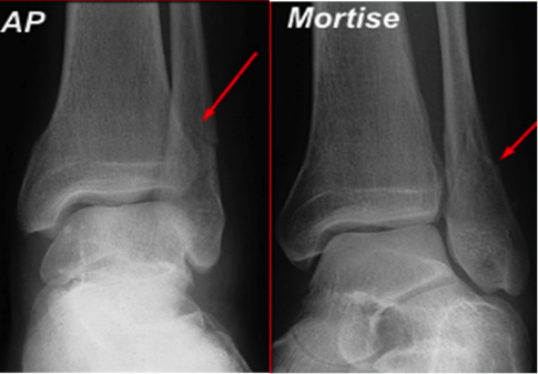
Isolated Posterior Malleolus Fracture
Picture
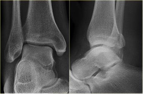
Bimalleolar fracture / equivalent
Picture
 
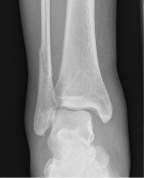
*Notice how there are two breaks to the ring structure, therefore this injury pattern is consistent with an unstable joint (increased medial clear space, distal fibula fracture). These unstable injuries convey a higher need for orthopedic intervention.
 
Associated syndesmotic injuries
​
Picture
Picture
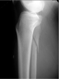
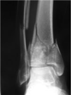
​Pilon Fracture
 
This following fracture pattern is often confused for an ankle fracture, however the presence of a pilon fracture conveys a different mechanism and prognosis.
 
A Pilon fracture, French for mortar and pestle, occurs due to an axial loading mechanism and is defined by articular impaction and comminution of the distal tibia. When you see a pilon fracture it is important to evaluate for other injuries typical of axial loading (i.e. lumbar spine, calcaneus, ect.)
Picture
Picture
Picture
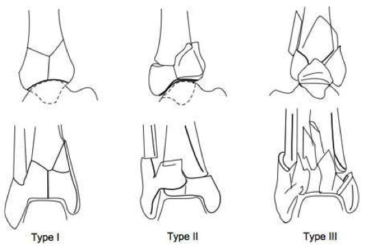
Sources
 
https://radiopaedia.org/articles/ankle-radiograph-an-approach
 
http://www.orthobullets.com/trauma/1047/ankle-fractures
 
http://www.orthobullets.com/trauma/1046/tibial-plafond-fractures?expandLeftMenu=true
 
https://radiopaedia.org/search?utf8=✓&q=pilon+fracture&scope=all
 
https://meds.queensu.ca/central/assets/modules/ts-ankle-radiograph/isolated_medial_malleolus_fracture.html
 

BY: DR. ETHEN ELLINGTON M.D.
2 Comments
Dhaneswar Bag link
5/4/2020 16:22:14

X-Ray Tech

Reply
Hasan Mahmoud Abdel kader link
7/15/2020 15:42:00

Ok

Reply

Your comment will be posted after it is approved.


Leave a Reply.

    Orthopedics Blog

    Picture

    Author 

    CMC ER Residents 
    (for learning purposes, imagine all cases present to a free standing ED for dispo and treatment options)

    Subscribe To Blog

    Archives

    June 2018
    April 2018
    March 2018
    October 2017
    September 2017
    February 2016
    January 2016
    December 2015
    November 2015
    October 2015
    September 2015
    August 2015
    July 2015
    June 2015
    May 2015
    April 2015
    March 2015
    February 2015
    January 2015
    December 2014
    November 2014

    Categories

    All
    Lower Extremity
    Nerve Injury
    Spine
    Sports Medicine
    Upper Extremity
    Vascular Injury

    RSS Feed

    Disclaimer: All images and x-rays included on this blog are the sole property of CMC EM Residency and cannot be used or reproduced without written permission.  Patient identifiers have been redacted/changed or patient consent has been obtained.  Information contained in this blog is the opinion of the author and application of material contained in this blog is at the discretion of the practitioner to verify for accuracy.
Powered by Create your own unique website with customizable templates.
  • RESIDENCY
    • About CMC
    • Curriculum
    • Benefits
    • Explore Charlotte
    • Official Site
  • FELLOWSHIP
    • EMS
    • Global EM
    • Pediatric EM
    • Toxicology >
      • Tox Faculty
      • Tox Application
    • (All Others)
  • PEOPLE
    • Program Leadership
    • PGY-3
    • PGY-2
    • PGY-1
    • Alumni
  • STUDENTS/APPLICANTS
    • Medical Students at CMC
    • EM Acting Internship
    • Healthcare Disparities Externship
    • Resident Mentorship
  • #FOAMed
    • EM GuideWire
    • CMC Imaging Mastery
    • Pediatric EM Morsels
    • Blogs, etc. >
      • CMC ECG Masters
      • Core Concepts
      • Cardiology Blog
      • Dr. Patel's Coding Blog
      • Global Health Blog
      • Ortho Blog
      • Pediatric Emergency Medicine
      • Tox Blog
  • Chiefs Corner
    • Top 20
    • Current Chiefs
    • Schedules >
      • Conference/Flashpoint
      • Block Schedule
      • ED Shift Schedule
      • AEC Moonlighting
      • Journal Club/OBP/Audits Schedule
      • Simulation
    • Resources >
      • Fox Reference Library
      • FlashPoint
      • Airway Lecture
      • Student Resources
      • PGY - 1
      • PGY - 2
      • PGY - 3
      • Simulation Reading
      • Resident Wellness
      • Resident Research
      • Resume Builder
    • Individualized Interactive Instruction