CMC COMPENDIUM
  • RESIDENCY
    • About CMC
    • Curriculum
    • Benefits
    • Explore Charlotte
    • Official Site
  • FELLOWSHIP
    • EMS
    • Global EM
    • Pediatric EM
    • Toxicology >
      • Tox Faculty
      • Tox Application
    • (All Others)
  • PEOPLE
    • Program Leadership
    • PGY-3
    • PGY-2
    • PGY-1
    • Alumni
  • STUDENTS/APPLICANTS
    • Prospective Visiting Students
    • UNC/Wake Forest Students
    • Healthcare Disparities Externship
    • Resident Mentorship
  • #FOAMed
    • EM GuideWire
    • CMC Imaging Mastery
    • Pediatric EM Morsels
    • Blogs, etc. >
      • CMC ECG Masters
      • Core Concepts
      • Cardiology Blog
      • Dr. Patel's Coding Blog
      • Global Health Blog
      • Ortho Blog
      • Pediatric Emergency Medicine
      • Tox Blog
  • Chiefs Corner
    • Top 20
    • Current Chiefs
    • Schedules >
      • Conference/Flashpoint
      • Block Schedule
      • ED Shift Schedule
      • AEC Moonlighting
      • Journal Club/OBP/Audits Schedule
      • Simulation
    • Resources >
      • Fox Reference Library
      • FlashPoint
      • Airway Lecture
      • Student Resources
      • PGY - 1
      • PGY - 2
      • PGY - 3
      • Simulation Reading
      • Resident Wellness
      • Resident Research
      • Resume Builder
    • Individualized Interactive Instruction

Hip Dislocations

11/25/2014

0 Comments

 
HPI: 
Patient 1: Restrained passenger of a head on MVC. 
Patient 2: Elderly patient with fall from standing.
 
Physical Exam: 
Patient 1: Hip flexed and internally rotated. Unable to straighten the leg. Unable to walk.
Patient 2: Hip flexed and externally rotated. Unable to straighten the leg. Unable to walk.
 
Imaging: 
Patient 1: Posterior hip dislocation of native hip. 
Patient 2: Superolateral hip dislocation of prosthetic hip. 

Posterior Hip Dislocation

Picture

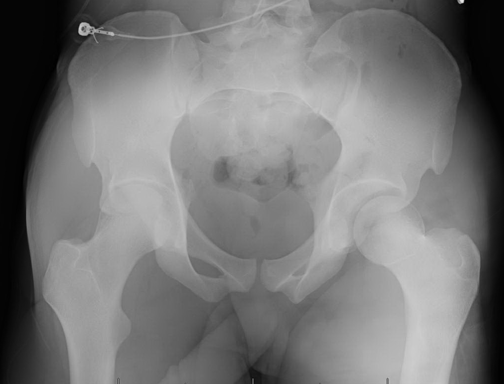
Prosthetic Hip Dislocation
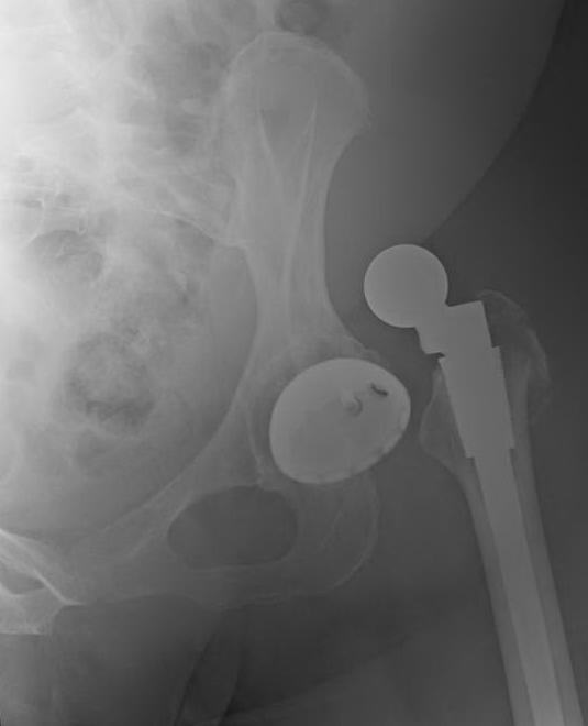
Picture
Treatment: 
  • Posterior hip dislocations: Stand on the bed over the patient, flex/internally rotate/adduct the hip and pull traction, then attempt to straighten out the leg while continuing traction.
 
  • Anterior hip dislocations: With leg extended, externally rotate and extend off the edge of the bed. Then pull traction and slowly internally rotate the leg while still in traction.
 
  • Native hip - Highest risk for femoral neck fractures w/ relocation. Emergently intubate, paralyze, and try to reduce as soon as possible (within 6 hours) before the muscles tighten down. Contraindication to reduction is a femoral neck or acetabular fracture whether displaced or non-displaced. Up to 20% chance of associated sciatic nerve injuries. Perform post-reduction CT bony pelvis for all traumatic hip dislocations of native hips.
 
  • Operative repair of native hip dislocation if... delayed presentation, femoral neck fracture, irreducible dislocation, or incarcerated bone fragment. Associated fractures of the acetabulum also require ORIF. 
 
  • Prosthetic hip - Patients go to the PACU w/ ortho for a propofol sedation and hip relocation. Discharged from the PACU if hip stable after relocation. No post-reduction CT required. 
 
  • 4-6 weeks of touch down weight bearing after native hip relocation.

****Don't forgot you can find the ED Policy for Deep Sedation on the Top 20 Page.  Anesthesia must be present for intubation.  Click here to read more.  
0 Comments

Your comment will be posted after it is approved.


Leave a Reply.

    Orthopedics Blog

    Picture

    Author 

    CMC ER Residents 
    (for learning purposes, imagine all cases present to a free standing ED for dispo and treatment options)

    Subscribe To Blog

    Archives

    June 2018
    April 2018
    March 2018
    October 2017
    September 2017
    February 2016
    January 2016
    December 2015
    November 2015
    October 2015
    September 2015
    August 2015
    July 2015
    June 2015
    May 2015
    April 2015
    March 2015
    February 2015
    January 2015
    December 2014
    November 2014

    Categories

    All
    Lower Extremity
    Nerve Injury
    Spine
    Sports Medicine
    Upper Extremity
    Vascular Injury

    RSS Feed

    Disclaimer: All images and x-rays included on this blog are the sole property of CMC EM Residency and cannot be used or reproduced without written permission.  Patient identifiers have been redacted/changed or patient consent has been obtained.  Information contained in this blog is the opinion of the author and application of material contained in this blog is at the discretion of the practitioner to verify for accuracy.
Powered by Create your own unique website with customizable templates.
  • RESIDENCY
    • About CMC
    • Curriculum
    • Benefits
    • Explore Charlotte
    • Official Site
  • FELLOWSHIP
    • EMS
    • Global EM
    • Pediatric EM
    • Toxicology >
      • Tox Faculty
      • Tox Application
    • (All Others)
  • PEOPLE
    • Program Leadership
    • PGY-3
    • PGY-2
    • PGY-1
    • Alumni
  • STUDENTS/APPLICANTS
    • Prospective Visiting Students
    • UNC/Wake Forest Students
    • Healthcare Disparities Externship
    • Resident Mentorship
  • #FOAMed
    • EM GuideWire
    • CMC Imaging Mastery
    • Pediatric EM Morsels
    • Blogs, etc. >
      • CMC ECG Masters
      • Core Concepts
      • Cardiology Blog
      • Dr. Patel's Coding Blog
      • Global Health Blog
      • Ortho Blog
      • Pediatric Emergency Medicine
      • Tox Blog
  • Chiefs Corner
    • Top 20
    • Current Chiefs
    • Schedules >
      • Conference/Flashpoint
      • Block Schedule
      • ED Shift Schedule
      • AEC Moonlighting
      • Journal Club/OBP/Audits Schedule
      • Simulation
    • Resources >
      • Fox Reference Library
      • FlashPoint
      • Airway Lecture
      • Student Resources
      • PGY - 1
      • PGY - 2
      • PGY - 3
      • Simulation Reading
      • Resident Wellness
      • Resident Research
      • Resume Builder
    • Individualized Interactive Instruction