|
Authored by: Blake Bauer, MD
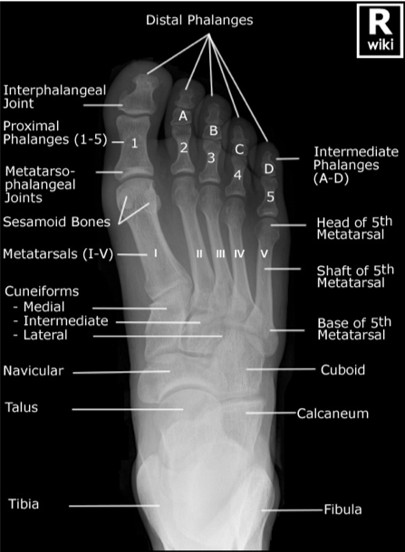
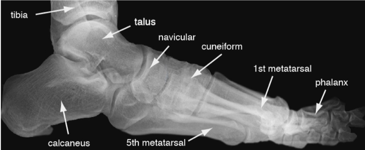
The Foot
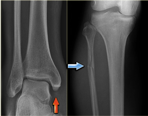
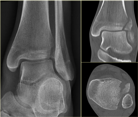
The Ankle and Lower Leg
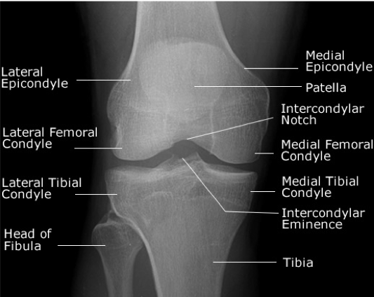
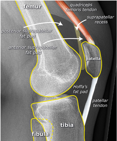
The Knee
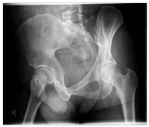
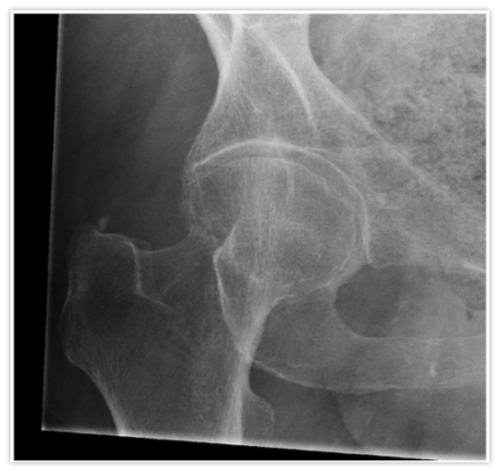
The Hip
1 Comment
5/16/2019 07:42:45
thankyou for informative blog. Dr.r. M Naveen Chandar Reddy, Orthopaedic Surgeon with around 10 years of experience.is the best doctor in hyderabad. <a href="https://https://www.drnaveenreddyortho.com//"><b>Best Joint Replacement Surgeon in Hyderabad</b></a> Your comment will be posted after it is approved.
Leave a Reply. |
Orthopedics Blog
AuthorCMC ER Residents Archives
June 2018
Categories
All
Disclaimer: All images and x-rays included on this blog are the sole property of CMC EM Residency and cannot be used or reproduced without written permission. Patient identifiers have been redacted/changed or patient consent has been obtained. Information contained in this blog is the opinion of the author and application of material contained in this blog is at the discretion of the practitioner to verify for accuracy.
|
 RSS Feed
