 Anatomy
- Internal carotid, sympathetic chain, IJ, cranial nerves - all pretty close in this small area Risks for PTA - smoking, bad teeth, chronic tonsillitis Complications - airway blockage, necrosis into carotid sheath, deep space infection, Lemierre's Syndrome Imaging Indications CT Scans should not be done routinely for PTA. PTA is primarily a clinical diagnosis. Consider Contrasted CT in: - toxic folks - immuncompromised - concern for deep space spread - Bilateral PTA (high extension rate) - uncertain diagnosis Ultrasound all PTA - Helps to improve diagnostic accuracy. - Helps to guide needle during aspiration. When to Consult - airway compromise - gas producing organism or air fluid levels - deep space infection - failure to respond in 48-72 hrs of IV anbx; - if meet indications for quinsy tonsillectomy (removal of tonsils while PTA is present) - Uncooperative patient - Pediatric patient - Recurrent tonsillitis or PTA - Severe trismus How to Drain 1. Apply anesthetic - 2 seconds of spray - hurricaine or cetacaine; atomize 4% lidocaine 2. Identify tonsil - use blood flow to help distinguish tonsil from abscess; Identify carotid 3. Supplies - need good lighting - bottom 1/2 of speculum or DL blade; - needle - 18 gauge only need 1 cm unsheathed; consider spinal needle 4. Aspiration - Parallel to floor of the mouth; start at superior pole of tonsil - if you don' t get pus - then move to middle, then third pole - this is the only way needle aspiration is comparable to I&D; - aspiration = less painful than I&D - speed of relief may be higher with I&D; ED literature rec's aspiration Medical Management 1. Antibiotics - group A strep and anaerobe coverage - Unasyn, Clindamycin, or Vancomycin (if life threatening or fail to respond) (everyone should get a dose of IV anbx) - Oral 10-14 day course Pen VK QID plus flagyl 500 QID; clindamycin 300-450mg q 6 hrs; augmentin 45 mg/kg q12 - in young folks test for mono 2. Steroids - jury is still out; initially decreases pain but in 48-72 hrs no difference; use IV solumedrol Disposition 1. 4-6 hrs observation, then DC if can tolerate PO; f/up in 24-48 hrs; gargle with H2O2 at least after each meal; soft diet and good oral hydration; antibiotics 2. Admit (23 hr obs on IV anbx) - peds; toxic; immuncompromised; can't tolerate PO
0 Comments
 Immune System is the Bouncer
- We know that there are bad microbes and good microbes. - It is now believed that the healthy Immune System acts much like a Bouncer at a Bar... allowing the good microbes in and keeping the bad microbes out. Illnesses associated with alterations in indigenous microbes - When "Bouncers go bad," illness like Obesity, IBD, Cdiff, asthma, or MRSA occur - Obesity - Giving antibx to animals make them gain weight - Obese people have less bacteroides - Markedly different gut flora in obese and skinny people - Inflammatory Bowel Disease - Increased correlation with antibiotic use - unclear causation - MRSA - Changes in microbiota related to MRSA status - Fecal transplants for recurrent C. difficile - Stool administered via NG tubes have a high success rate *Theoretically antibiotics in the last 70 yrs have altered normal human microbes*  Electrical Storm
- Refractory Ventricular rhythm , >3 episodes in 24 hours - Fix the underlying causes (ischemia, electrolytes, arrythmogenic meds) - LBBB & MI - difficult to interpret - remember your Sgarbossa Criteria! - Amiodarone is medication of choice, add beta blockade early with refractory condition Ectopic Pregnancy - Ectopic pregnancy - number 1 cause of 1st trimester deaths - Goal of the ED provider - EXCLUDE ECTOPIC by confirming IUP! - Start scan by finding the uterus! - Endomyometrial mantle - distance from the wall of the gestational sac to the outer wall of the uterus - in a normal pregnancy, measurement should be >0.8 cm - Switch to transvaginal probe if unable to obtain adequate views - IUP can only be diagnosed by yolk sac or fetal pole within gestational sac 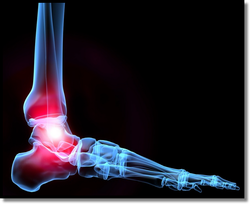 The Ankle
- 3 Bones - 3 Primary Joints - Medial mal with medial talus - Tibial plafond with talar dome - Lateral mal with lat talus Ankle Xrays - Medial clear space --- if over 4mm, its concerning for syndesmotic injury or possible deltoid ligament incompetence - Tib-fib clear space --- greater than 5mm is abnormal - Bi-mal and Tri-mal fxs often need surgery - Pilon fractures ---- high injury mechanism!! - Be sure to get additional films to look at tibia, knee and also LUMBAR SPINE - Pediatric patients are different - Ligaments stronger - More likely to fracture bone than sprain ligament The Foot - Foot has 28 bones - Divided into hindfoot, midfoot and forefoot - Hindfoot controls inversion and eversion - Midfoot controls foot abduction and adduction - Forefoot controls plantarflexion and dorsiflexion - Calcaneal fracture --- 10% associated lumbar fractures!! - Talar neck fractures - very bad injury, minimal blood flow there; often times need OR for reduction - LisFranc fracture dislocation - axial load and foot planted. - Can be severe or subtle. - If concerned for subtle, then get full weight bearing films. - Jones fx - 5th metatarsal head fracture - Put in post-op shoe or splint, follow up; Majority non-op  GU & Pelvic Trauma Basics
Bladder injury - Pelvic fracture, direct blow, penetrating injury - bladder rupture in 5-10% of pelvic fractures (more fractures = higher risk of injury) - Intraperitoneal - dome is the weakest part (surgical repair) - Extraperitoneal - manage with foley Evaluation of the GU Tract
- Look for extravasation & look for contrast in bladder - if no contrast in the bladder = complete disruption of urethra - this requires surgery; some partial injuries can be managed with Foley catheter.
Upper Tract Injury - flank bank or abdominal pain with gross hematuria or microscopic hematuria with shock - 85% of renal injuries are secondary to blunt trauma; - any penetrating trauma near this area requires evaluation of kidneys - management depends on grade of injury - Ureteral injuries - easy to miss; tend to present late Pelvic Ring Fractures - pelvis is strong - takes a lot of force to break it; Assess for injuries: a. Proximate - urethra, bladder, vagina, sciatic nerve b. Distant - brain, chest, aorta, intra-abdominal Who to image- physical findings suggestive of injury, shock Types: 1. Lateral compression - horizontal anterior ring fracture- look for sacral fracture 2. AP - open book fracture - high risk of bleeding out - "mac daddy of pelvic fracture" 3. Vertical Sheer - high risk of vascular injury - if no femur fracture put in traction 4. Posterior ring disruption - increased mortality Unstable patients with pelvic fracture - angio vs OR: if grossly positive FAST, OR first. If not, angio first.  Central Cord Syndrome
-Generally caused by hyperextension. Patients with history of central canal stenosis at risk -Upper extremities affected > lower extremities -Distal affected > proximal -Usually bladder dysfunction Opioid Induced Hearing Loss -Occurs <72 hours after use (seen in both acute and chronic users) -MCC is hydrocodone and heroin but seeing more with methadone lately -May be unilateral or bilateral -Most resolve within 72 hours but may be permanent -Treat with cessation of narcotics and possibly cochlear implants if permanent Rhabdomyolysis -Fluids, fluids, fluids as treatment -Diuretics (mannitol) and bicarb are controversial -Risk of AKI is lower when CK <5,000 but can be seen at CK levels of 1,000 -Urine dipstick + for blood with urinalysis - for blood has sensitivity of 80% for diagnosis  Retroperitoneal Organs
Physical Exam Findings
Symptoms 1. Most common presenting complaint = abdominal pain; then leg/hip pain; back pain 2. Can have femoral neuropathy, iliopsoas spasm Traumatic RPH
Spontaneous RPH
Management - controversial Zone I - concern for vascular injury - likely OR Zone II & III - ?pulsatile, ?expanding - determines intervention Packing vs arterial embolization - majority of traumatic RPH are venous in nature CORE CONCEPTS: - RPH is a rare diagnosis with significant mortality - see keep on your differential! - Undress your adult patients too - look for Fox's sign, etc - Most common presenting symptom: abdominal pain, then leg or back pain - Seen more commonly in elderly and those on anticoagulation, but 1/3 of pts who presented with spontaneous RPH were not on anticoagulation  Heart Lesions
1. Left to right - VSD, ASD, cushion defect, PDA 2. Cyanotic - truncus, transposition, total anomalous, tricuspid atresia, tetralogy a. Cyanosis - decrease of deoxygenated hgb by 3-5 mg/dl 1. Shunting from lung 2. Mixing blue and red blood 3. Single ventricle Break the left side of the heart (Hypoplastic left, aortic stenosis, coarct) --> hepatomegaly, gray, pulmonary edema, etc Break the right side of the heart (hypoplastic right heart, tricupsid atresia, pulmonary atresia, tetrology of fallot) --> Blue, poor perfusion, acidosis Not all ductal dependent lesions are cyanotic - AS, coarct Not all cyanotic lesions are ductal dependent - truncus arteriosis, TAPR Cyanotic Lesions a. Truncus Arteriosus - Blue because they're mixing - mixing happens before duct, therefore not ductal dependent - Pulmonary exam will vary; You can give them O2 - won't worsen cyanosis but won't help b. Transposition of Great Arteries - Cyanotic because you have mixing blood; If you find these later (ie, in the ED and not immediately after birth) these kids will all have VSD; Ductal dependent O2 wont help but wont hurt c. Tricuspid Atresia - Blue because not perfusing lungs; Right ventricle doesn't develop (Hypoplastic right heart) Ductal dependent; only pulmonary artery flow will come through ductus from aorta ECG will show LVH but only because right side isn't balancing it out O2 will prob not help, but won't kill d. Tetrology of Fallot - Cyanotic because of decreased pulmonary perfusion and mixing - O2 can help e. Totally Anomalous Pulmonary Venous Return - Cyanotic because of mixing Hyperoxia Test - 10 minutes of 100% O2 and see response > may help differentiate between pulmonary and cardiac etiology * For cyanotic lesions oxygen is not going to kill - it just may not help* * O2 can hurt you on left to right shunts* Left to right shunts are usually dyspneic/hypoxic because they are over-perfusing the lungs and they get fluid overload. Oxygen will cause vasodilitation of the pulmonary vessels and increase left to right shunting worsening the problem. Prostaglandins - 0.05-0.1 mcg/kg/min > will cause apnea - tube the kid  Myocardial Infarction in TTP
Penetrating Scrotal Trauma:
Diagnotic Error in Medical Decision Making
 Mechanical Issues
Acute mitral regurg - 7 to 10 days post MI - can result from papillary muscle rupture - often complication of inferior MI - can cause abrupt cardiogenic shock - best tx = afterload reduction with diuretics, nitroprusside, Surgery. Ventricular septal rupture - first 24 hrs and at 2-5 days - findings: holosystolic murmur, CP, painless hypotension - tx: O2, surgery, vasodilators, nitroprusside Ventricular aneurysm - findings: CHF +/- shock - dx: CXR w/ enlarged silhouette - At risk for thromboembolism. Ventricular free wall rupture - cause hemopericardium/tamponade - signs: CP, hypotension, tamponade Post MI Arrhythmias
Dressler's Syndrome
|
Archives
August 2018
Categories
All
|
 RSS Feed
