CMC COMPENDIUM
  • RESIDENCY
    • About CMC
    • Curriculum
    • Benefits
    • Explore Charlotte
    • Official Site
  • FELLOWSHIP
    • Global EM
    • Toxicology
    • (All Others)
  • PEOPLE
    • Program Leadership
    • PGY-3
    • PGY-2
    • PGY-1
    • MATCH 2022
    • Alumni
  • STUDENTS/APPLICANTS
    • Prospective Visiting Students
    • Healthcare Disparities Externship
    • UNC/Wake Forest Students
  • #FOAMed
    • EM GuideWire
    • CMC Imaging Mastery
    • Pediatric EM Morsels
    • Blogs, etc. >
      • CMC ECG Masters
      • Core Concepts
      • Cardiology Blog
      • Dr. Patel's Coding Blog
      • Global Health Blog
      • Ortho Blog
      • Pediatric Emergency Medicine
      • Tox Blog
  • Chiefs Corner
    • Top 20
    • Current Chiefs
    • Schedules >
      • Conference/Flashpoint
      • Block Schedule
      • ED Shift Schedule
      • AEC Moonlighting
      • Journal Club/OBP/Audits Schedule
      • Simulation
    • Resources >
      • Fox Reference Library
      • FlashPoint
      • Airway Lecture
      • Student Resources
      • PGY - 1
      • PGY - 2
      • PGY - 3
      • Simulation Reading
      • Resident Wellness
      • Resident Research
      • Resume Builder
    • Individualized Interactive Instruction
    • Evaluations/Interview Season

The significance of Respiratory artifact (RA) in the ECG - Dr. Littmann

5/5/2016

0 Comments

 
Picture
I.  Characteristics of respiratory artifact
  1. Repetitive microoscillations, best seen in the inferolateral leads; rarely seen in leads I and V1
  2. Occasionally, the microoscillations are preceded by what look like P waves - the presence of pseudo-P waves may mimic atrial dissociation
  3. The RA corresponds to the inspiratory phase of the respiratory cycle
  4. Duration of the RA corresponds to duration of the inspiratory phase of respiration
 
II.  The clinical significance of respiratory artifact
  1. The presence of RA always indicates increased work of breathing, respiratory distress
  2. It allows precise measurement and tracking of the respiratory rate - count the number of RAs in the 10-sec 12-lead ECG and multiply it by 6; measure 1 or more respiratory cycles in mm and divide it into 1,500 or its multiples
  3. 44% of pts with RA required ventilation support; 28% required intubation, mechanical ventilation
  4. Successful treatment of the underlying condition resulted in a decreased rate or elimination of RA
 
III.  Respiratory artifact can aid in the recognition of sleep-disordered breathing
  1. Central sleep apnea is characterized by
                a. marked sinus deceleration or AV block followed by
                b. an abrupt increase in heart rate and the
                c. simultaneous appearance of very fast RA
  1. Obstructive sleep apnea is characterized by
                a. profound sinus bradycardia with
                b. simultaneously occurring very long respiratory (snoring) artifact followed by
                c. a sudden increase in the heart rate and resolution of the RA
  1. Cheyne-Stokes breathing is characterized by
                 a. periodic, cyclic clustering of RA
                 b. the onset of most periods which contain the RAs show the characteristics of central sleep apnea
​


0 Comments

Sharpen YOur Calipers - Dr. Littmann

1/28/2016

0 Comments

 
Picture
What is STEMI without STE?
  1. Patients presenting with acute chest pain may have clinical STEMI (acute total thrombotic occlusion of a large coronary artery) but no obvious ST-segment elevation in the ECG
  2. This occurs with STEMI locations that are not well represented by conventional ECG leads
  3. Such patients are at high for not receiving timely reperfusion therapy and for having poor outcomes
  4. Easily memorizable ECG signs can help raise awareness of the possibility of STEMI without STE
  5. If suspected, placing exploring electrodes to the area of interest can quickly uncover the STEMI
 
Posterolateral or High Posterior MI
  1. The most common type of missed STEMI
  2. Almost always due to acute occlusion of the LCX
  3. May involve the posterior papillary muscle and can cause severe mitral regurgitation
  4. Earliest ECG sign: ST depression in the anterior chest leads (mirror image of posterior ST elevation)
  5. Frequent associated findings: subtle Q waves or subtle ST elevation in the inferior or lateral leads
  6. Can be uncovered by placing exploring electrodes to the back (V7-V8-V9)
 
High Lateral MI
  1. The second most commonly missed STEMI
  2. Almost always due to acute occlusion of the first diagonal branch of the LAD (LAD-D1)
  3. Fortunately usually small
  4. Earliest ECG sign: ST elevation in leads I, aVL and V2, but not in V1 or V3, and ST depression in III and aVF
  5. With the conventional 4x3 lead display format, the spacing of ST deviation resembles the shape of the South African flag (the “South African Flag Sign”)
  6. Can be uncovered by placing the V4-V5-V6 ECG leads 1 and 2 interspaces higher
 
RV infarct
  1. Almost always due to acute occlusion of the proximal RCA
  2. Almost always associated with acute inferior STEMI
  3. Recognition of RV infarct, therefore, is now less important because code STEM is usually called anyway for the inferior MI
  4. Recognition can be important, however, if the ST elevation in the inferior leads is subtle or nondiagnostic
  5. Clues to suspect RV MI:
    1. Inferior STEMI with marked ST depression in I and aVL (left leads)
    2. Inferior STEMI with ST elevation in V1 only but not in V2
  6. Can be uncovered by placing right-sided chest leads
  7. If inferior STEMI has been diagnosed, there is no need to waste time to record right-sided chest leads
  8. Suspected RV MI:
    1. Use caution with vasodilators (nitrates)
    2. Consider IV fluids for hypotension with JVD

0 Comments

Runs of Wide-Complex Tachycardia on Telemetry - Dr. Littmann

11/12/2015

0 Comments

 
Picture
Differential Diagnosis
  1. SVT with aberrancy (SVT with RBBB or SVT with LBBB)
  2. Nonsustained VT
  3. Artifact
  4. Uncertain (“I don’t know”) – this is a better choice than incorrect guessing!
 
When the intrinsic rhythm is sinus:
I. What was the first beat of the tachycardia?
  1. If the first early beat was a PAC (premature P wave in front of the QRS) -> SVT
  2. If the first early beat was a PVC (no premature P wave in front of the QRS) -> VT
II. What is the P-QRS relationship?
  1. A-V dissociation present -> VT
  2. More QRS complexes than P waves -> VT
III. Fusion complexes present? -> VT
  1. Combination QRS morphology (QRS wider than during sinus but narrower than the widest QRS)
  2. P waves must be present in front of the combination morphology QRS complexes
 
When the intrinsic rhythm is atrial fibrillation:
  1. If the WCT is regular -> VT
 
Artifact
Always consider artifact if:
  1. It is uncertain which was the first or the last beat of the WCT
  2. The rate of the tachycardia was excessive
  3. The patient remained asymptomatic during a long run of a very fast tachycardia
Prove artifact by demonstrating that the
  1. Intrinsic QRS complexes march through
  2. “Notches” march through
  3. Simultaneous hemodynamic tracing indicates regularity

0 Comments

Sharpen Your Calipers: Wide Complex Tachycardia - Dr. Littmann

9/10/2015

0 Comments

 
Picture
General
1.     WCT algorithms do not work in the real life setting
2.     Don’t pay attention to what others have said; you need to evaluate the ECG yourself
3.     First question: is the WCT regular or irregular?

Sustained Regular WCT
I. Differential Diagnosis
1.     Sustained regular WCT: most likely ventricular tachycardia (VT)
a.     Spend 2 minutes searching for P waves – inconsistent P-QRS relationship is 100% specific for VT
b.     Bizarre QRS morphology that is not c/w RBBB or LBBB (negative QRS in lead I; QRS concordance in the chest leads) strongly supports VT

2.     Consider SVT if QRS morphology resembles RBBB or LBBB
a.     Find an old ECG: are the QRS morphologies identical?
b.     If no old ECG available, prove SVT by giving IV adenosine
c.     “I think this is SVT” is not good enough!

II. Therapy of Sustained Ventricular Tachycardia
1.     Unstable: electric cardioversion

2.     Clinically stable: one round of IV antiarrhythmic drug therapy allowed
          a.     IV procainamide may be the most effective agent (avoid in patients with severe systolic CHF and 

                   in patients with baseline prolonged QT)
          b.     IV amiodarone
          c.     Consider IV propranolol or IV metoprolol in ischemic VT and in patients with “electric storm” 

                  (repeat episodes of VT/VF)

Sustained Irregular WCT

I. Differential Diagnosis
1.     Sustained irregular WCT: most likely not VT but atrial fibrillation (AF)
2.     Rate 120-160; QRS morphology is c/w RBBB or LBBB: AF with bundle branch block
3.     Rate very fast; QRS morphology is bizarre, not c/w RBBB or LBBB: most likely AF with WPW (FBI – Fast, Broad-complex, Irregular)

II. Therapy
1.     AF with BBB: usual therapy for AF (IV diltiazem etc.)
2.     AF with WPW (“FBI”)
          a.     Clinically unstable or ventricular rate excessive: electric cardioversion
          b.     Clinically stable: IV procainamide or IV ibutilide
          c.     Clinically very stable, rate not very fast: PO flecainide or propafenone
NOT ALLOWED: IV verapamil, diltiazem, digoxin, adenosine

Picture
0 Comments

Sharpen Your Calipers - Dr. Littmann

8/20/2015

0 Comments


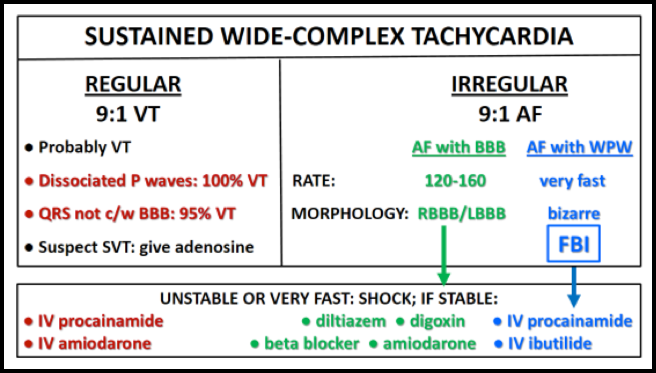
Bifascicular Block and Second Degree AV Block

1.     In asymptomatic individuals, chronic bifascicular block does not usually require cardiac work-up; the prognosis is generally benign

2.     The following high-risk features, however, warrant urgent evaluation:
           a.   Bifascicular block and syncope
           b.   Bifascicular block and intermittent second degree AV block
           c.   1:1 AV conduction at slower sinus rates but higher grade block (i.e., 2:1 AV conduction) at faster 

                  sinus rates (“acceleration-dependent AV block”)

3.     Evaluation and management of patients with bifascicular block:
          a.   Actively search for nonconducted P waves in the 12-lead ECG
          b.   Always review telemetry strips and actively search for episodes of second degree AV block      

                 (blocked P waves)
          c.   In symptomatic patients with bifascicular block who develop acceleration-dependent second 

                 degree AV block and a very slow ventricular rate, carotid massage or IV beta blocker can 
                 paradoxically restore 1:1 AV conduction
          d.   Patients with bifascicular block and syncope require admission and cardiology consultation for 

                 possible pacemaker implantation
          e.   Patients with bifascicular block and intermittent second degree AV block require cardiology 

                 consultation for possible pacemaker implantation

The Pacemaker ECG

1.     Ventricular pacing: always try to determine what the atria are doing

2.     Sinus P wave in front of each paced QRS complex indicates dual chamber (A-V sequential) pacemaker where the ventricular pacer is tracking sinus rhythm

3.     Two pacer spikes about 5 mm apart indicate AV sequential pacing

4.     If there are no P waves or 2 pacer spikes, search for the presence of retrograde P waves after the paced QRS complexes; retrograde P waves are sharp negative in the inferior leads (in II, III and aVF) and usually upright in V1

5.     If there are no P waves in front of the paced QRS complexes and no retrograde P waves present, always consider the possibility of underlying atrial fibrillation

0 Comments

    Archives

    August 2018
    February 2018
    January 2018
    December 2017
    October 2017
    September 2017
    August 2017
    July 2017
    June 2017
    May 2017
    April 2017
    March 2017
    February 2017
    January 2017
    December 2016
    November 2016
    October 2016
    September 2016
    August 2016
    July 2016
    June 2016
    May 2016
    April 2016
    March 2016
    February 2016
    January 2016
    December 2015
    November 2015
    October 2015
    September 2015
    August 2015
    July 2015
    June 2015
    May 2015
    April 2015
    March 2015
    February 2015
    January 2015
    December 2014
    November 2014
    October 2014
    September 2014
    August 2014
    July 2014
    June 2014
    May 2014
    April 2014
    March 2014
    February 2014
    January 2014
    December 2013
    November 2013
    October 2013
    September 2013
    August 2013
    July 2013

    Categories

    All
    Abdominal Pain
    Abdominal-pain
    Airway
    Back Pain
    Back Pain
    Bleeding
    Change-in-mental-status
    Chest Pain
    Dizziness
    Ecg
    Emboli
    Environmental
    Fever
    Gyn
    Headache
    Hypertension
    Infectious Disease
    Pain
    Pediatric Emergency
    Professionalism
    Psych
    Respiratory Distress
    Sepsis
    Shock
    Toxins
    Trauma
    Vomiting
    Weakness

    RSS Feed

    Tweets by @PedEMMorsels
Disclaimer: All original material and images included on this website are the sole property of CMC EM Residency and cannot be used or reproduced without written permission.  Information contained on this website is the opinion of the authors and does not necessarily represent the official opinion of Atrium Health or Carolinas Emergency Medicine Residency. 

For Health Care Providers:  Every effort is made to provide the most up to date evidence based medicine.  However, this content may not necessarily reflect the standard of care and application of material contained on this website is at the discretion of the practitioner to verify for accuracy.

For the Public:  This content is not intended to be a substitute for professional medical advice, diagnosis, or treatment.  Relying on information contained on this website is done at your own risk.  Do not disregard professional medical advice or delay seeking care secondary to content on this website.  Call 911 or seek a medical professional immediately for any medical emergencies.
Like us on Facebook or
follow us on Twitter/Instagram

Contact Us:

Department of Emergency Medicine
Medical Education Building., Third floor
1000 Blythe Blvd.
Charlotte, NC 28203

Telephone: 704-355-3658 
Fax: 704-355-7047
​
  • RESIDENCY
    • About CMC
    • Curriculum
    • Benefits
    • Explore Charlotte
    • Official Site
  • FELLOWSHIP
    • Global EM
    • Toxicology
    • (All Others)
  • PEOPLE
    • Program Leadership
    • PGY-3
    • PGY-2
    • PGY-1
    • MATCH 2022
    • Alumni
  • STUDENTS/APPLICANTS
    • Prospective Visiting Students
    • Healthcare Disparities Externship
    • UNC/Wake Forest Students
  • #FOAMed
    • EM GuideWire
    • CMC Imaging Mastery
    • Pediatric EM Morsels
    • Blogs, etc. >
      • CMC ECG Masters
      • Core Concepts
      • Cardiology Blog
      • Dr. Patel's Coding Blog
      • Global Health Blog
      • Ortho Blog
      • Pediatric Emergency Medicine
      • Tox Blog
  • Chiefs Corner
    • Top 20
    • Current Chiefs
    • Schedules >
      • Conference/Flashpoint
      • Block Schedule
      • ED Shift Schedule
      • AEC Moonlighting
      • Journal Club/OBP/Audits Schedule
      • Simulation
    • Resources >
      • Fox Reference Library
      • FlashPoint
      • Airway Lecture
      • Student Resources
      • PGY - 1
      • PGY - 2
      • PGY - 3
      • Simulation Reading
      • Resident Wellness
      • Resident Research
      • Resume Builder
    • Individualized Interactive Instruction
    • Evaluations/Interview Season