CMC COMPENDIUM
  • RESIDENCY
    • About CMC
    • Curriculum
    • Benefits
    • Explore Charlotte
    • Official Site
  • FELLOWSHIP
    • EMS
    • Global EM
    • Pediatric EM
    • Toxicology >
      • Tox Faculty
      • Tox Application
    • (All Others)
  • PEOPLE
    • Program Leadership
    • PGY-3
    • PGY-2
    • PGY-1
    • Alumni
  • STUDENTS/APPLICANTS
    • Medical Students at CMC
    • EM Acting Internship
    • Healthcare Disparities Externship
    • Resident Mentorship
  • #FOAMed
    • EM GuideWire
    • CMC Imaging Mastery
    • Pediatric EM Morsels
    • Blogs, etc. >
      • CMC ECG Masters
      • Core Concepts
      • Cardiology Blog
      • Dr. Patel's Coding Blog
      • Global Health Blog
      • Ortho Blog
      • Pediatric Emergency Medicine
      • Tox Blog
  • Chiefs Corner
    • Top 20
    • Current Chiefs
    • Schedules >
      • Conference/Flashpoint
      • Block Schedule
      • ED Shift Schedule
      • AEC Moonlighting
      • Journal Club/OBP/Audits Schedule
      • Simulation
    • Resources >
      • Fox Reference Library
      • FlashPoint
      • Airway Lecture
      • Student Resources
      • PGY - 1
      • PGY - 2
      • PGY - 3
      • Simulation Reading
      • Resident Wellness
      • Resident Research
      • Resume Builder
    • Individualized Interactive Instruction

OrthoClass - Pediatric Elbow - Dr. S. Lawson

7/28/2017

0 Comments

 
Picture
The basics of evaluation for elbow fractures in children are the same as adults - 2 (ideally 3) views, determine if there are any disruptions in the cortex, assess the radiocapitellar and anterior humeral lines (both should bisect the capitellum), and look for signs of hemarthrosis (anterior or lateral fat pads).

Most elbow fractures in children are extension type supracondylar fractures. The Gartland classification system can be used to describe fractures:
  1. Type I - minimal to no displacement with no disruption in the cortex
  2. Type II - displacement with an interrupted anterior cortex but intact posterior cortex
  3. Type III - displaced with disruption of both the anterior and posterior cortex

The elbow is not fully developed until late adolescence. The timing of ossification center development varies from child to child, but girls generally develop slightly earlier than boys. The order of appearance IS reliable - use mnemonic "CRITOE" to remember the order:
  • Capitellum
  • Radial head
  • Internal (medial) epicondyle
  • Trochlea
  • Olecranon
  • External (lateral) epicondyle.

Knowing the order of development can help you determine if that small piece of bone you're seeing is an ossification center or a bone fragment (comparison films of the contralateral elbow may prove helpful as well).
A thorough neurovascular exam is the most important part of your assessment and will help determine management. For a quick motor exam:
  • thumbs up checks the radial nerve
  • fingers in a "fat five" and crossing the index and middle finger for "good luck" checks the ulnar nerve
  • a true "okay sign" (flexion at the DIP of thumb and index finger) checks the median nerve, specifically the anterior interosseous (AIN) branch, which is the most commonly injured in supracondylar fractures
For a quick sensation exam:
  • dorsal first webspace tests the radial nerve
  • palmar aspect of the index finger tests the median nerve
  • palmar aspect of the pinky finger tests the ulnar nerve

Check a radial pulse, and assess color, temperature, and CAP REFILL. Pink and pulseless can be okay; pale and pulseless, however, is not.

If you can't feel a pulse, listen for triphasic or biphasic Doppler flow.

Type I fractures can be splinted in a posterior long arm splint with close Ortho follow-up
Type II fractures can be managed like a type I if there is minimal displacement and swelling and the neurovascular exam is normal - otherwise splint and arrange urgent operative repair
Type III will always require operative repair - emergently if a poor neurovascular exam, urgently if a normal exam


0 Comments

Management of Atrial Fibrillation in the Acutely Ill

7/27/2017

0 Comments

 
Picture
  • In the acutely ill patient with rapid Afib, management should focus on quick rate control
  • IV diltiazem should be the treatment of choice
  • Bolus is crucial; re-bolus is crucial – they brake the vicious cycle of fast ventricular rate, reduced LV filling, reduced stroke volume, all resulting in further adrenergic activation
  • Diltiazem drip does not reduce the heart rate; it maintains whatever you achieved with the bolus
  • Never order “diltiazem drip, titrate to heart rate”
  • Ideal dose of initial diltiazem bolus is 0.25 mg/kg over 2 minutes
  • Ideal dose of re-bolus is 0.35 mg/kg over 2 minutes
  • Marked hypotension may limit giving the optimum doses of diltiazem boluses
  • Under these circumstances, there are several options for pre-treatment before the IV diltiazem bolus
    • IV fluid boluses
    • IV digoxin, 0.5 mg
    • Phenylephrine, 100-300 mcg IV push over 10-30 sec
  • With the help of one or more of the above, IV diltiazem can almost always be given safely
  • Consider IV beta blocker (esmolol) instead of diltiazem for patients with rapid Afib and acute MI, ongoing ischemia, thyrotoxicosis and HOCM crisis
  • IV amiodarone is almost never needed for rate control
  • If rate control is still inadequate despite maximum tolerated doses of diltiazem, adding 0.5 mg of IV digoxin will frequently “finish the job”
  • For critically ill patients with rapid Afib, consider emergent cardioversion
  • Do not shock the patient with MAT or with repetitive Afib characterized by the presence of occasional sinus complexes
  • These patients are frequently good candidates for rhythm management with amiodarone or beta blocker


0 Comments

Carolinas Case Conference

7/27/2017

0 Comments

 
Picture
1. ACS in the elderly can be tricky. Presentations can be subtle with the most common symptoms being chest pain, shortness of breath, diaphoresis and nausea. Patients > 65 who present without chest pain who have ACS have significantly higher mortality.

2. When making a diagnosis of DVT/PE consider why this patient is hyper-coagulable. Seek common etiologies and refer or admit for further workup if necessary.
 
3. Consider vascular surgery consult for large DVTs in proximal vessels in patient's with low risk for bleeding complications, as these patients may benefit from catheter directed thrombolysis or thrombectomy.
 
4. Signs of traumatic aortic injury include: widened mediastinum, abnormal contour of aorta, depressed left mainstem bronchus, deviation of NGT to right, loss of aortopulmonary window.


0 Comments

Leadership in the Trauma Bay

7/21/2017

0 Comments

 
Picture
Before the Code
  • Consider
    • How busy is department?
    • Staffing?
    • Number of patients?
Always wear gown, gloves, masks, shoe covers, lead gown and thyroid shield.
Recognize shock!
  • Consider:
  • Vital Signs
  • Character of pulses
  • Diaphoresis
  • Skin Temp
  • Mental Status
  • Lactate, base deficit
Pain Management
  • Fentanyl
  • Ketamine
  • US guided nerve blocks
Set priorities and control consultants
 
CT scan: A Dark and Lonely Place Where Trauma Patients Go to Die.
     - Be sure about your patient before they leave your side and go to the doughnut.


0 Comments

Pre-PICU Prep - Dr. Sean Fox

7/21/2017

0 Comments

 
Picture
Shock: Recognition
  • There is no single pathognomonic finding that defines shock.
  • Hypotension is a late finding, but an ominous one, in kids.
  • Constellation of findings:
  1. Tachycardia
  2. Tachypnea
  3. Poor perfusion
  4. Poor pulse quality
  5. Altered mental status
  • Cold Shock findings:
  1. High Systemic Vascular Resistance
  2. Cold, clammy, mottled, or cyanotic extremities
  3. Capillary Refill > 2 seconds
  4. Diminished / thready pulses
  5. Narrow pulse pressure.
  • Respect the “just ain’t right” findings:
  1. Poor feeding
  2. Jittery
  3. Irritable
  4. Lethargic 
Be Aggressive Early
  • Once recognized, be aggressive within 1st hour!
  • IV or IO 40-60 ml/kg of isotonic fluids PUSHED rapidly
  1. Do not hang to gravity or on a “pump.”
  2. Use syringe pushes or pressure bags
  3. Children commonly will require 40-60 ml/kg in the 1st hour, but may require more (some say 200 ml/kg in 1st hour in right clinical setting). 
  • Don’t forget about Glucose! 
  • Optimize oxygenation
  1. Supplemental may be all that is initially needed.
  2. 30-40% of a child’s cardiac output goes to the work of breathing when critically ill, so often will require additional support (i.e., intubation).
  • Broad spectrum antibiotics
Fluid-Refractory Shock
  • Keep your Differential open!
    • While ordering empiric antibiotics, consider the other causes of SHOCK in children.
    • The child with fluid-refractory shock deserves a second and third consideration for the other possible culprits!
  • Use your bedside Ultrasound
  1. Pericardial Effusion & Tamponade?
  2. Overview of heart function / squeeze / size
  3. IVC volume? – perhaps more fluids aren’t the answer
  4. Pneumothorax?
  5. Free intra-abdominal fluid? – Is there occult trauma??
Vasopressors can be Started Peripherally
  • Do not hesitate to start vasopressors.
    • Children with fluid-refractory shock tend to respond to inotropes. 
    • Reversing shock is associated with better survival.
  • Common perception is that vasoactive medications (vasopressors) need to be give via central line.
    • In an ideal setting, this is reasonable. That 1st hour of critical illness is often not ideal.
    • There is no data clarifying whether one vasopressor is more harmful when given peripherally than another. 
  • Epinephrine has been shown to be safe and effective when given via peripheral IV or IO in the setting of Septic Shock.  [Ramaswamy, 2016; Ventura, 2015]
  • Time is critical; central lines aren’t easy in children; PIVs and IOs work just fine! 


0 Comments

Carolinas Case Conference - Dr. J. Raper

7/20/2017

0 Comments

 
Picture
Submassive Pulmonary Embolism
  • a nefarious and cryptic disease
  • definition in our shop:
    • lobar or greater clot burden
    • normotension
    • Right ventricular dilatation/dysfunction by CT,  cardiac biomarker, or echocardiogram
  • In normotensive patients who are found to have RVD:
    • Risk of shock is 10%
    • Risk of death is 5%
  • In patients who receive TPA for PE:
    • Risk of major hemorrhage is 24%
    • Risk of hemorrhagic stroke is 3-5%
  • So who gets consideration for lytics (again, in our shop)?
    • Submassive PE with:
      • Elevated shock index
      • Episodic hypotension
      • SaO2 <92% with distress
    • All Massive PE (Lobar or proximal clot with sustained hypotension or bradycardia)

Acute Retroviral Syndrome
  • Phase during acute infection when HIV RNA and p24 are detectable but Ab is not
  • Viral loads can reach >10,000,000 copies
  • High rates of transmission to intimate partners
  • 80-98% are symptomatic
  • onset is 1-4 weeks following exposure
  • Symptoms include:
    • Fever
    • Myalgias
    • Skin rash
    • Headache
  • When to test?
    • Suspicion for another STD
    • Opportunistic infection
    • High risk sexual behavior
    • Signs of  acute retroviral syndrome
  • Early identification benefits during acute infection include:
    • Reduction of transmission to partners
    • Reduction of viral diversity, which reduces resistance
    • Reduction of lymphatic latent infection/inflammation
    • Prevention of delay in diagnosis


0 Comments

Syphilis - Dr. M. Noe

7/13/2017

0 Comments

 
Picture
  1. Syphilis is caused by an old bacterium (spirochaete bacterium Treponema pallidum) but is making a comeback, with infection rates greater than HIV in the US. 
  2. Various stages of disease affect many organ systems:
    1. Primary – painless chancre, local adenopathy
    2. Secondary – diffuse non-pruritic rash, flu symptoms, condyloma lata
    3. Tertiary – neuro, cardiac, gummas
  3. Testing: VDRL/RPR for screening & follow up, FTA-ABS/TP-PA for confirmation
  4. Penicillin is your friend! – Benzithine penicillin remains the treatment of choice for all stages of syphilis infection, though duration of treatment varies depending on stage.
  5. Knowledge of syphilis as a disease came at a price, teaching us a valuable lesson about the role that physicians play in their patients' lives and the incredible trust they place in the profession.

0 Comments

Carolinas case conference - Dr. S. Pecevich

7/13/2017

0 Comments

 
Picture
Internal Hernias
  • With patients status post gastric bypass, beware of the internal hernia and have low threshold to consult gastric bypass surgeon in abdominal pain!
  • CT scan may fail to reveal internal hernia in up to 20% of cases, and the diagnosis can be missed on initial read.
  • Abdominal pain in pregnancy? There’s more than just the uterus, so remain vigilant and keep a broad differential.
  • Be wary of sending people home after repeat IV pain medication.
 
Osteomyelitis, MSSA bacteremia and pediatrics
  • No great single test to diagnose osteomyelitis--keep a high index of suspicion.
  • In pediatrics, lactate can be unreliable and should be used to rule in, not rule out.
  • Pediatric populations can decompensate quickly, and a normal BP is likely the last vital sign to go in setting of shock. Track perfusion, as skin mottling, AMS and urine output will change before the patient’s BP will.
  • MSSA toxic shock — PVL is a toxin seen in MSSA osteomyelitis with high morbidity.
  • Vancomycin should be used as initial antibiotic when concerned for staph bacteremia. Vancomycin over Clindamycin -- May be up to 30% MRSA clindamycin resistance in Charlotte area,

0 Comments

Idiopathic Intracranial Hypertension - Dr. A. Dozois

7/13/2017

0 Comments

 
Picture
  1. In the right clinical context, decreased visual acuity + papilledema = BAD. These patients need workup including LP and MRI in the ED, and urgent referral to ophthalmology (for optic nerve sheath fenestration) or neurosurgery (for shunt placement).
  2. Papilledema can be difficult to recognize. Beware of mimics such as optic nerve head drusen and decreased optic disc height. 
  3. LP is essential in the workup of IIH, but is limited by many potential confounders. To maximize accuracy in obtaining an opening pressure reading, remember la clé (French for "the KEY")- Calm patient, Lateral decubitus position, knees Extended

0 Comments

LVAD EMERGENCIES - Dr. Erin Noste

7/13/2017

0 Comments

 
Picture
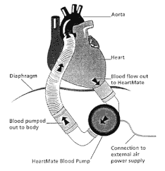
 LVAD patient in ED = evaluate for (LVAD)2
 
L = Look, listen, and feel the LVAD device
- Look: Connection & controller, green light with no alarm = good sign
- Listen: For the hum of the LVAD, a quiet LVAD is BAD news
- Feel: Hot LVAD controller = BAD, could mean distal obstruction, dislodgment, thrombosis 
 
V = Venous Thromboembolism, Ventricle
- Venous Thromboembolism at greatest risk when INR < 1.5 with increased risk of TIA/Stroke and PE/DVT
- Pump Thrombosis Signs = LVAD is hot, working hard, high RPM, low flow, dilated RV/LV, Low MAP 
- Right Ventricle: Signs of Right Heart Strain, RV failure is VERY concerning for LVAD patients, consider right sided STEMI, PE
 
A = Anti-Coagulation, Arrhythmias 
- Anti-Coagulation: Bleeding risk from anti-coagulation and acquired von Willebrand syndrome (increased shear stress and decrease pulsatility of LVAD) = GI bleeds and head bleeds 
- Arrhythmias: check EKG on LVAD patients, can be in VF and still awake and talking, safe to defibrillate LVAD patient (if not emergent with consultation of LVAD team) 
 
D = Drive Line, Dehydration 
- Drive Line Infections: 40-60% of patients will develop an infection, also consider a deep pocket infection and obtain imaging to identify source of infection
- Dehydration: Very common in patients as they adjust to not having to be fluid restrictive, leading to increased risk of suction events and arrhythmias, LVADs love FLUID


0 Comments
<<Previous

    Archives

    August 2018
    February 2018
    January 2018
    December 2017
    October 2017
    September 2017
    August 2017
    July 2017
    June 2017
    May 2017
    April 2017
    March 2017
    February 2017
    January 2017
    December 2016
    November 2016
    October 2016
    September 2016
    August 2016
    July 2016
    June 2016
    May 2016
    April 2016
    March 2016
    February 2016
    January 2016
    December 2015
    November 2015
    October 2015
    September 2015
    August 2015
    July 2015
    June 2015
    May 2015
    April 2015
    March 2015
    February 2015
    January 2015
    December 2014
    November 2014
    October 2014
    September 2014
    August 2014
    July 2014
    June 2014
    May 2014
    April 2014
    March 2014
    February 2014
    January 2014
    December 2013
    November 2013
    October 2013
    September 2013
    August 2013
    July 2013

    Categories

    All
    Abdominal Pain
    Abdominal-pain
    Airway
    Back Pain
    Back Pain
    Bleeding
    Change-in-mental-status
    Chest Pain
    Dizziness
    Ecg
    Emboli
    Environmental
    Fever
    Gyn
    Headache
    Hypertension
    Infectious Disease
    Pain
    Pediatric Emergency
    Professionalism
    Psych
    Respiratory Distress
    Sepsis
    Shock
    Toxins
    Trauma
    Vomiting
    Weakness

    RSS Feed

    Tweets by @PedEMMorsels
Powered by Create your own unique website with customizable templates.
  • RESIDENCY
    • About CMC
    • Curriculum
    • Benefits
    • Explore Charlotte
    • Official Site
  • FELLOWSHIP
    • EMS
    • Global EM
    • Pediatric EM
    • Toxicology >
      • Tox Faculty
      • Tox Application
    • (All Others)
  • PEOPLE
    • Program Leadership
    • PGY-3
    • PGY-2
    • PGY-1
    • Alumni
  • STUDENTS/APPLICANTS
    • Medical Students at CMC
    • EM Acting Internship
    • Healthcare Disparities Externship
    • Resident Mentorship
  • #FOAMed
    • EM GuideWire
    • CMC Imaging Mastery
    • Pediatric EM Morsels
    • Blogs, etc. >
      • CMC ECG Masters
      • Core Concepts
      • Cardiology Blog
      • Dr. Patel's Coding Blog
      • Global Health Blog
      • Ortho Blog
      • Pediatric Emergency Medicine
      • Tox Blog
  • Chiefs Corner
    • Top 20
    • Current Chiefs
    • Schedules >
      • Conference/Flashpoint
      • Block Schedule
      • ED Shift Schedule
      • AEC Moonlighting
      • Journal Club/OBP/Audits Schedule
      • Simulation
    • Resources >
      • Fox Reference Library
      • FlashPoint
      • Airway Lecture
      • Student Resources
      • PGY - 1
      • PGY - 2
      • PGY - 3
      • Simulation Reading
      • Resident Wellness
      • Resident Research
      • Resume Builder
    • Individualized Interactive Instruction