CMC COMPENDIUM
  • RESIDENCY
    • About CMC
    • Curriculum
    • Benefits
    • Explore Charlotte
    • Official Site
  • FELLOWSHIP
    • EMS
    • Global EM
    • Pediatric EM
    • Toxicology >
      • Tox Faculty
      • Tox Application
    • (All Others)
  • PEOPLE
    • Program Leadership
    • PGY-3
    • PGY-2
    • PGY-1
    • Alumni
  • STUDENTS/APPLICANTS
    • Prospective Visiting Students
    • UNC/Wake Forest Students
    • Healthcare Disparities Externship
    • Resident Mentorship
  • #FOAMed
    • EM GuideWire
    • CMC Imaging Mastery
    • Pediatric EM Morsels
    • Blogs, etc. >
      • CMC ECG Masters
      • Core Concepts
      • Cardiology Blog
      • Dr. Patel's Coding Blog
      • Global Health Blog
      • Ortho Blog
      • Pediatric Emergency Medicine
      • Tox Blog
  • Chiefs Corner
    • Top 20
    • Current Chiefs
    • Schedules >
      • Conference/Flashpoint
      • Block Schedule
      • ED Shift Schedule
      • AEC Moonlighting
      • Journal Club/OBP/Audits Schedule
      • Simulation
    • Resources >
      • Fox Reference Library
      • FlashPoint
      • Airway Lecture
      • Student Resources
      • PGY - 1
      • PGY - 2
      • PGY - 3
      • Simulation Reading
      • Resident Wellness
      • Resident Research
      • Resume Builder
    • Individualized Interactive Instruction

M&M - Dr. Bronner

9/7/2013

0 Comments

 
Case 1 -    Post-Operative Pneumoperitoneum and Perforated Marginal Ulcer After Roux-en-Y

  1. Pneumoperitoneum, or "Free air"
    1. 90% of the time this is perforated viscous until proven otherwise!
    2. CT far more sensitive in detecting than plain films.
    3. ONLY CONSIDER NON-SURGICAL IF:
      1. No fever
      2. No Abdominal Pain
      3. No Leukocytosis
      4. No Peritonitis

    4. For the other 10%:
      1. MOST COMMON cause is Post-Surgical Pneumoperitoneum (PP)
        1. Will be present in 44% of post-op patients in 3 days
        2. 30% on days 4-18
        3. Should be resolved in all patients in 18 days
        4. Drains increase PP rate more than double (52% vs 21%)
      2. Other causes:
        1. Pseudopneumoperitoneum - Radiographic Artifact
        2. Thoracic sources - CPR, PTX, Mechanical Ventilation
        3. Gyn sources - Procedures, Vaginal Insufflation
        4. Misc - Cocaine, SCUBA, Idiopathic

  2. Complications of Bariatric Surgery  (i.e. CALL BLUE SURGERY EARLY!!!)
    1. Understand major types and their complications
      1. Review Articles worth your time:
        1. Campanile FC, Boru CE, Rizzello M, Puzziello A, Copaescu C, Cavallaro G, Silecchia G. Acute complications after laparoscopic bariatric procedures: an update for the general surgeon.
        2. Langenbecks Arch Surg. 2013;398:669-686.Edwards ED, Jacob BP, Gagner M, Pomp A. Presentation and Management of Common Post-Weight Loss Surgery Problems in the Emergency Department. Ann Emerg Med. 2006;47:160-166.
      2. Can be found here! (click link!)
      3. Gibbs Bonus factoid! -- Gastric banding on xray -
        1. 45 degree rectangle = normal
        2. Horizontal = band migration and needs deflation


Case 2 - Complications of Chronic Suppurative Otitis Media (CSOM)
  1. Ruptured TM from Otitis - Quinolone drops are initial therapy, no aminoglycocides (ototoxic)
  2. TM rupture over 6 weeks = ENT Consult!
  3. CSOM with mastoid tenderness - CT brain +/- temporal bone
    1. Also be VERY CONCERNED if fever, AMS, N/V, focal neuro deficits
  4. Intratemporal and Intracranial Infections need IV Ab
    1. Cover for Staph (incl MRSA), Pseudomonas, Hemophilus PLUS Enteric bugs via eustachian tubes
    2. FIRST LINE OPTION:  Cefepime, Flagyl, Vanc, +/- otic quinolone
  5. Great review of the badness transmitted through the temporal bones found here!
    1. Smith JA, Danner CJ. Complications of Chronic Otitis Media and Cholesteatoma. Otolaryngol Clin N Am. 2006;39:1237-1255.
  6. ADDITIONAL DISCUSSION POINTS:
    1. LP without CT is safe (NEJM 2001, 345: 1727-33)
      1. No Papilledema
      2. Age < 60
      3. No immmunocompromised
      4. No hx of CNS disease
      5. No seizure within 1 week
      6. Physical Exam - normal, non focal with normal mental status
    2. When not to do LP on CT findings
      1. Midline shift
      2. Obstructive hydrocephalus
      3. Basilar cistern compression
      4. Posterior fossa mass (displacement or compression of 4th ventricle)
    3. Gibbs factoid - Herniation after LP is likely Urban Legend

Case 3 - Acetabular Protrusio

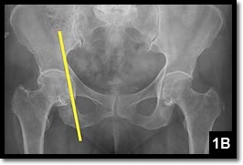
Important Radiographic Signs to Identify on AP Pelvis/Hip
        Ilioischial Line (Kohler's Line)
  1. Straight Line along medial border of Ilium through Ischium (Inf Pubic Ramus)
  2. If femoral head bulges inside of this line = acetabular disruption        


        Center Edge Angle of Wiberg
      1. Find center of femoral head
      2. Draw vertical line from this point
      3. Draw second line from this point to roof of acetabulum
        1. 20-40 degrees = Normal
        2. <20 degrees = Suspect Developmental Hip Dysplasia
        3. >40 degrees = Acetabular Protrusion
Picture
Picture
0 Comments

Your comment will be posted after it is approved.


Leave a Reply.

    Archives

    August 2018
    February 2018
    January 2018
    December 2017
    October 2017
    September 2017
    August 2017
    July 2017
    June 2017
    May 2017
    April 2017
    March 2017
    February 2017
    January 2017
    December 2016
    November 2016
    October 2016
    September 2016
    August 2016
    July 2016
    June 2016
    May 2016
    April 2016
    March 2016
    February 2016
    January 2016
    December 2015
    November 2015
    October 2015
    September 2015
    August 2015
    July 2015
    June 2015
    May 2015
    April 2015
    March 2015
    February 2015
    January 2015
    December 2014
    November 2014
    October 2014
    September 2014
    August 2014
    July 2014
    June 2014
    May 2014
    April 2014
    March 2014
    February 2014
    January 2014
    December 2013
    November 2013
    October 2013
    September 2013
    August 2013
    July 2013

    Categories

    All
    Abdominal Pain
    Abdominal-pain
    Airway
    Back Pain
    Back Pain
    Bleeding
    Change-in-mental-status
    Chest Pain
    Dizziness
    Ecg
    Emboli
    Environmental
    Fever
    Gyn
    Headache
    Hypertension
    Infectious Disease
    Pain
    Pediatric Emergency
    Professionalism
    Psych
    Respiratory Distress
    Sepsis
    Shock
    Toxins
    Trauma
    Vomiting
    Weakness

    RSS Feed

    Tweets by @PedEMMorsels
Powered by Create your own unique website with customizable templates.
  • RESIDENCY
    • About CMC
    • Curriculum
    • Benefits
    • Explore Charlotte
    • Official Site
  • FELLOWSHIP
    • EMS
    • Global EM
    • Pediatric EM
    • Toxicology >
      • Tox Faculty
      • Tox Application
    • (All Others)
  • PEOPLE
    • Program Leadership
    • PGY-3
    • PGY-2
    • PGY-1
    • Alumni
  • STUDENTS/APPLICANTS
    • Prospective Visiting Students
    • UNC/Wake Forest Students
    • Healthcare Disparities Externship
    • Resident Mentorship
  • #FOAMed
    • EM GuideWire
    • CMC Imaging Mastery
    • Pediatric EM Morsels
    • Blogs, etc. >
      • CMC ECG Masters
      • Core Concepts
      • Cardiology Blog
      • Dr. Patel's Coding Blog
      • Global Health Blog
      • Ortho Blog
      • Pediatric Emergency Medicine
      • Tox Blog
  • Chiefs Corner
    • Top 20
    • Current Chiefs
    • Schedules >
      • Conference/Flashpoint
      • Block Schedule
      • ED Shift Schedule
      • AEC Moonlighting
      • Journal Club/OBP/Audits Schedule
      • Simulation
    • Resources >
      • Fox Reference Library
      • FlashPoint
      • Airway Lecture
      • Student Resources
      • PGY - 1
      • PGY - 2
      • PGY - 3
      • Simulation Reading
      • Resident Wellness
      • Resident Research
      • Resume Builder
    • Individualized Interactive Instruction