CMC COMPENDIUM
  • RESIDENCY
    • About CMC
    • Curriculum
    • Benefits
    • Explore Charlotte
    • Official Site
  • FELLOWSHIP
    • EMS
    • Global EM
    • Pediatric EM
    • Toxicology >
      • Tox Faculty
      • Tox Application
    • (All Others)
  • PEOPLE
    • Program Leadership
    • PGY-3
    • PGY-2
    • PGY-1
    • Alumni
  • STUDENTS/APPLICANTS
    • Medical Students at CMC
    • EM Acting Internship
    • Healthcare Disparities Externship
    • Resident Mentorship
  • #FOAMed
    • EM GuideWire
    • CMC Imaging Mastery
    • Pediatric EM Morsels
    • Blogs, etc. >
      • CMC ECG Masters
      • Core Concepts
      • Cardiology Blog
      • Dr. Patel's Coding Blog
      • Global Health Blog
      • Ortho Blog
      • Pediatric Emergency Medicine
      • Tox Blog
  • Chiefs Corner
    • Top 20
    • Current Chiefs
    • Schedules >
      • Conference/Flashpoint
      • Block Schedule
      • ED Shift Schedule
      • AEC Moonlighting
      • Journal Club/OBP/Audits Schedule
      • Simulation
    • Resources >
      • Fox Reference Library
      • FlashPoint
      • Airway Lecture
      • Student Resources
      • PGY - 1
      • PGY - 2
      • PGY - 3
      • Simulation Reading
      • Resident Wellness
      • Resident Research
      • Resume Builder
    • Individualized Interactive Instruction

Upper GI Bleed in Pediatrics - Dr. Walker

6/8/2017

0 Comments

 
1.  Signs and symptoms of an Upper GI bleed in the pediatric patient
            -most common presentation is hematemesis
            -melena also common presentation 
             -many things mimic GI bleed -> food coloring, raw meats, swallowed blood from oropharynx 

2.  Differential diagnosis for acute pediatric upper GI bleed
        -determine if variceal vs nonvariceal 
         -variceal bleed (uncommon but can be life threatening)-> portal HTN from congenital liver pathologies 
         -mucosal bleed -> gastritis, esophagitis, caustic ingestion, foreign body most common in pediatrics 

3.  ED diagnostic workup for an upper GI bleed in a pediatric patient 
       -CBC, BMP, LFT's, and Type/Cross essential to the workup

4. ED Management of Pediatric Upper GI bleed patient 
       -Place an NG tube 
       -Get GI, Surgery, and Interventional Radiology Involved early 
       -Octreotide and vasopressin are important treatments for variceal bleed 
      -For mucosal bleeds control acid production ​
0 Comments

Scary Neonate Case COnference - Dr. Bryant

4/20/2017

0 Comments

 
Picture
- Neonate in respiratory distress: remember NRP!
- Neonate in respiratory distress with true unilateral absent breath sounds = Congenital Diaphragmatic Hernia??!!-> confirm with CXR

Congenital Diaphragmatic Hernia (CDH)
- Neonates likely have high risk for pulmonary hypertension!
- Do not give PPV -> worsens GI distention/lung compression = worsening pulmonary HTN
- In neonates intubate early to prevent hypoxia (hypoxia worsens pulmonary HTN) with low pressure vent settings
- Maintain systemic BP to reduce right to left shunting
- Older children with CDH, less likely to have significant pulmonary HTN, keep calm and try to avoid intubation
​

- Persistent/worsening cyanotic neonate - think cyanotic congenital heart defect -> Start Prostaglandins
- Remember Prostaglandins cause apnea, will likely need intubation
- Neonate in extremis = call for back-up early!

​


Picture
0 Comments

June 02nd, 2016

6/2/2016

0 Comments

 
Picture
  • ​URIs are common in children... acute sinusitis is not.  Approximately 6-8% of children with URI symptoms meet criteria for acute bacterial sinusitis
  • Diagnostic criteria for acute bacterial sinusitis have been revised in the last few years.  There are three clinical courses that constitute a diagnosis acute bacterial sinusitis...
                 - persistent symptoms without improvement
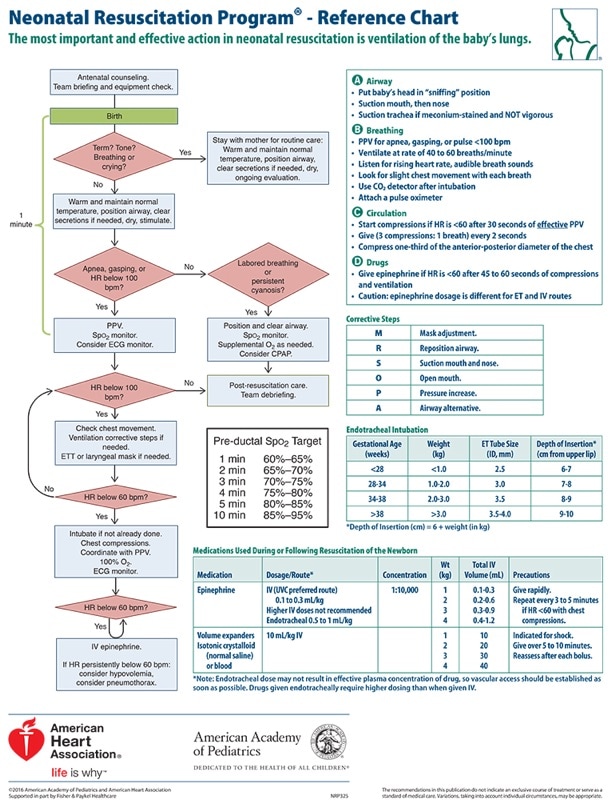
                 - severe onset of symptoms
                 - worsening clinical course

  • So that means, we don't diagnose sinusitis in children with imaging studies.
  • However, if you are worried about orbital or CNS involvement, it is recommended to evaluate with contrast CT or MRI.
  • It is most often caused by S. pneumoniae, H. influenzae, and M. catarrhalis.  S. aureushas not been identified as a major etiology.

  • Antibiotic treatment should take into account S. pneumoniae resistance patterns and beta lactamase production of H. influenzae and M. catarrhalis - high dose amoxicillin (90mg/kg/day BID) or Augmentin (90mg amoxicillin/kg/day BID) are first line choices.

0 Comments

Pediatric Case COnference - Dr. Lawson

4/26/2016

0 Comments

 
Picture
Abdominal Pain of Unusual Cause
  • Abdominal pain in children is not always AGE, appendicitis, or UTI - keep your differential broad and use your exam skills (including GU and Tanner staging)
  • "Smokiness" on ultrasound may be due to blood (RBCs scattering sound waves) or stasis
  • Hematocolpos as a result of imperforate hymen can cause abdominal pain and urinary retention

PE vs Sepsis
  • PE is a rare occurrence in children and often presents atypically (classic symptoms often only present in large PEs)
  • Adult clinical decision rules (Wells Criteria and PERC) do not reliably apply to children
  • Obesity, OCP use, and previous thrombus (not PE) are the three leading risk factors for PE
  • History of central venous line is the most important predisposing cause of DVT
  • Several studies other than CTA can help lessen your suspicion for PE (CXR, EKG, cardiac echo, extremity DVT US, BNP, troponin)
  • Sepsis in children can be subtle and precipitous

0 Comments

Pediatric ECGs - Dr. Bryant

4/21/2016

0 Comments

 
Picture

  • Normal Peds EKG Variations
    • Right heart dominance 
      • From infancy to adulthood ventricular dominance transitions from a relatively thicker RV in utero/infancy to thicker LV as adults
      • Right axis deviation 
      • Tall R waves in V1-V3
      • RSR' in V1
      • Deep S waves and Q waves in lateral leads
      • Juvenile T wave pattern in V1-V3
    • Small cardiac size = shorter intervals, faster rate
    • J-point depression
    • Early repolarization
    • Sinus arrhythmia
​
  • NOT Normal Peds EKG Variations
    • Brugada Syndrome
      • EKG: Leads V1-V3
        • RSR’ with ST elevation
      • 3 Types
        • Type 1 – coved
          • Diagnostic
        • Type 2/3 – saddle-back
    • Arrhythmogenic Right Ventricular Dysplasia (ARVD)
      • EKG: Leads V1-V3
        • Epsilon waves
        • Inverted T waves
    • Catecholaminergic Paroxysmal Ventricular Tachycardia (CPVT)
      • EKG:
        • Normal baseline, bidirectional QRS polymorphic Vtach with exertion/excitement
    • Hypertrophic Obstructive Cardiomyopathy (HOCM)
      • EKG: Lateral leads
        • Dagger-like deep narrow Q waves
        • Deep narrow T wave inversions
        • +/- LVH, left axis deviation
    • Wolff-Parkinson-White (WPW)
      • EKG:
        • Delta waves
        • Wide QRS
        • Short PQ segment
    • Anomalous Left Coronary Artery from the Pulmonary Artery (ALCAPA)
      • EKG:
        • Anterolateral ischemic changes
        • Lateral leads
          • Deep wide Q waves
          • T wave inversions
    • Long QT Syndrome
      • EKG:
        • QTc > 450ms
        • At risk for Torsade de Pointes/Vtach

0 Comments

Pediatric Syncope - Dr. Bryant

4/21/2016

0 Comments

 
Pediatric Syncope Algorithm
0 Comments

VP Shunts - Dr. Angela Johnson

2/18/2016

0 Comments

 
Picture
  • Be suspicious of shunt problems in …. Kids with shunts
  • HA, nausea, vomiting, irritability, behavior change are the weaker predictors you may miss if not vigilant
  • Malfunction common early, but eventually almost ALL get revised
  • Infection most common in first six months
  • Look at your own images!
  • Not stable – Call NSGY and consider tap


0 Comments

Pediatric Airway - Dr. Goode

2/11/2016

0 Comments

 
Picture
• The pediatric airway has known unique challenges. Prepare yourself physically and cognitively!

• Maintain spontaneous ventilation if there is any doubt about obtaining successful advanced airway.
​

• Practice using airway equipment when you don't need it, so you are ready when you do need it
.


0 Comments

CMC Case COnference - Dr. West

12/10/2015

0 Comments

 
Picture
Septic arthritis of the hip
-Most common hematongenous spread
- Up to 20% of patients with non-gonococcal septic arthritis will have 2 or more joints involved –> always do a full musculoskeletal exam!
- 50% will have positive blood cultures –> always obtain cultures.
- Risk factors: Extremes of age, hardware/recent instrumentation, skin infection, underlying arthritis, IV drug use
- You cannot rule out septic arthritis with inflammatory markers or any physical exam findings, so err on the side of obtaining joint fluid.
 
Pediatric septic arthritis vs. transient synovitis
- Kocher criteria can help differentiate: Temperature >38.5, WBC >12K, ESR >40, unable to bear weight.
  • 0/4: <0.2%, 1/4: 3%, 2/4: 40%, ¾: 93%, 4/4: 99.6
  • Note: this was a retrospective study and external validation studies did not perform as well.
  • Can not be used if patient recently on antibiotics.
- There is a MSK screening MRI protocol if you are concerned about LE deep space infection but having difficulty localizing the joint
 
Contrast Extravasation
  • Monitor area for signs of compartment syndrome or airway compromise (depending on location)
  • Complications rare now that we routinely use low-osmolar nonionic contrast
 
Spontaneous Pneumomediastnum
  • Under recognized cause of chest pain
  • Similar risk factors as spontaneous pneumothorax (asthma, tall, thin, valsalva, intense sporting activities)
  • Alveolar ruptures into surrounding bronchovascular sheath and free air tracks into mediastinum
  • Rarely causes tension physiology
  • If history concerning for esophageal pathology, consider CT esophagram
  • Treat conservatively by avoiding valsalva and barotrauma 
​

0 Comments

TEAM Case: Ped Status Epilepticus - Dr. Magill

12/3/2015

0 Comments

 
Picture
​Define status epilepticus:
  • >5 min seizure- impending status epilepticus
  • >30 min- established SE
  • >60 min- refractory SE

Consider etiologies:
  • Trauma/bleed
  • AVM Malformation
  • Febrile seizures
  • Infection
  • Tumor
  • Subtherapeutic meds
  • Lowered threshold with infection
  • Stroke
  • Cardiac/arrhythmia
  • Hypertensive Crisis
  • Pyridoxine deficiency/INH overdose
  • NMDA receptor Ab
  • FIRES

Remember ABCs and supportive care in addition to treating seizures

Learn dosing for hypoglycemic seizure with dextrose
  • Rule of 50's

Learn dosing for hyponatremia seizure with 3% NaCl 

Medications
First line:
  • Midazolam 0.15 mg/kg IV, 0.3 mg/kg buccal
  • Lorazepam 0.05-0.1 mg/kg IV, max 4 mg/dose rpt x 1
  • Diazepam 0.05-0.3 mg/kg IV, 0.5 mg/kg PR, max 5 mg

Second line:
  • Fospheny load 15-20 mg/kg IV
  • Levetiracetam load 15-20 mg/kg IV
  • Valproate load 20-40 mg/kg IV

Refractory/Third line:
  • Phenobarb load 20 mg/kg IV, 1 mg/kg/min
  • Pentobarb 5 mg/kg load
  • Propofol 2.5-3.5 mg/kg IV, rate 0.1-0.3 mg/kg/min
  • Ketamine 0.5-2 mg/kg IV, rate 5-20 mcg/kg/min
  • Pyridoxine 70 mg/kg max 5g, repeat as needed

0 Comments
<<Previous

    Archives

    August 2018
    February 2018
    January 2018
    December 2017
    October 2017
    September 2017
    August 2017
    July 2017
    June 2017
    May 2017
    April 2017
    March 2017
    February 2017
    January 2017
    December 2016
    November 2016
    October 2016
    September 2016
    August 2016
    July 2016
    June 2016
    May 2016
    April 2016
    March 2016
    February 2016
    January 2016
    December 2015
    November 2015
    October 2015
    September 2015
    August 2015
    July 2015
    June 2015
    May 2015
    April 2015
    March 2015
    February 2015
    January 2015
    December 2014
    November 2014
    October 2014
    September 2014
    August 2014
    July 2014
    June 2014
    May 2014
    April 2014
    March 2014
    February 2014
    January 2014
    December 2013
    November 2013
    October 2013
    September 2013
    August 2013
    July 2013

    Categories

    All
    Abdominal Pain
    Abdominal-pain
    Airway
    Back Pain
    Back Pain
    Bleeding
    Change-in-mental-status
    Chest Pain
    Dizziness
    Ecg
    Emboli
    Environmental
    Fever
    Gyn
    Headache
    Hypertension
    Infectious Disease
    Pain
    Pediatric Emergency
    Professionalism
    Psych
    Respiratory Distress
    Sepsis
    Shock
    Toxins
    Trauma
    Vomiting
    Weakness

    RSS Feed

    Tweets by @PedEMMorsels
Powered by Create your own unique website with customizable templates.
  • RESIDENCY
    • About CMC
    • Curriculum
    • Benefits
    • Explore Charlotte
    • Official Site
  • FELLOWSHIP
    • EMS
    • Global EM
    • Pediatric EM
    • Toxicology >
      • Tox Faculty
      • Tox Application
    • (All Others)
  • PEOPLE
    • Program Leadership
    • PGY-3
    • PGY-2
    • PGY-1
    • Alumni
  • STUDENTS/APPLICANTS
    • Medical Students at CMC
    • EM Acting Internship
    • Healthcare Disparities Externship
    • Resident Mentorship
  • #FOAMed
    • EM GuideWire
    • CMC Imaging Mastery
    • Pediatric EM Morsels
    • Blogs, etc. >
      • CMC ECG Masters
      • Core Concepts
      • Cardiology Blog
      • Dr. Patel's Coding Blog
      • Global Health Blog
      • Ortho Blog
      • Pediatric Emergency Medicine
      • Tox Blog
  • Chiefs Corner
    • Top 20
    • Current Chiefs
    • Schedules >
      • Conference/Flashpoint
      • Block Schedule
      • ED Shift Schedule
      • AEC Moonlighting
      • Journal Club/OBP/Audits Schedule
      • Simulation
    • Resources >
      • Fox Reference Library
      • FlashPoint
      • Airway Lecture
      • Student Resources
      • PGY - 1
      • PGY - 2
      • PGY - 3
      • Simulation Reading
      • Resident Wellness
      • Resident Research
      • Resume Builder
    • Individualized Interactive Instruction