CMC COMPENDIUM
  • Prospective Applicants
    • 2020 MATCH
    • About CMC
    • Our Curriculum
    • Our Residents
    • Our Fellowships
    • Program Leadership
    • Explore Charlotte
    • Official Site
  • ROTATING STUDENTS
    • Prospective Visiting Students
    • UNC Students
    • Healthcare Disparities Externship
    • Current Students
  • Current Residents
    • Airway Lecture!
    • PGY - 1
    • PGY - 2
    • PGY - 3
    • Simulation Reading
    • Blogs >
      • EM GuideWire
      • CMC ECG Masters
      • Core Concepts
      • #FOAMed
      • Cardiology Blog
      • Dr. Patel's Coding Blog
      • Global Health Blog
      • Ortho Blog
      • Pediatric Emergency Medicine
      • Tox Blog
    • Board Review
    • Journal Club
    • Resident Wellness
    • Resident Research
  • Top 20
  • Chiefs Corner
    • Schedules >
      • Conference/Flashpoint
      • Block Schedule
      • ED Shift Schedule
      • AEC Moonlighting
      • Journal Club/OBP/Audits Schedule
      • Simulation
    • Individualized Interactive Instruction
    • Evaluations/Interview Season
    • Contact Info
    • Resume Builder

Putting The K in EKG

8/31/2015

0 Comments

 
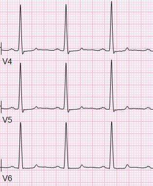
HPI: 
Patient is a 50 yo F with a history of ESRD s/p failed renal transplant now on hemodialysis who had onset of GI bleeding with associated lightheadedness and hypotension. An EKG was obtained. 

EKG:
Picture
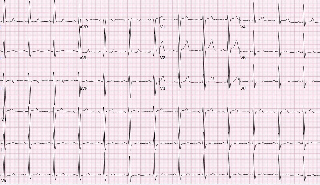
Question: 
What finding on this EKG warrants further management? 

EKG Interpretation: 
Normal sinus rhythm at a rate of 77, normal PR interval, normal QRS, normal axis, LVH (significantly increased voltages), narrow based, peaked T-waves, T-wave inversions in leads III and aVF. 

Discussion:
The narrow based, peaked T-waves present in this patient’s EKG, most evident in leads V3-V4, indicate probable hyperkalemia in this patient who is at risk for elevated potassium given her ESRD. The T-wave abnormalities in this EKG are somewhat subtle, but the narrow, “pinched down” bases of the T-waves should warn you of hyperkalemia. This patient’s potassium level was found to be 6.9 and was appropriately treated.

Pro-tip: 
  • Lead V4 can be your best friend for visualizing a lot of pathology on EKGs, including hyperkalemia, so make sure you pay attention to that upper right corner for subtle signs of elevated potassium! 
  • Remember to compare to an old EKG if available. 
  • Post treatment EKG shows much smaller, more typical broad based T waves:
Picture
Potassium can be the great imitator on EKGs because it can cause a wide variety of abnormal EKG findings, including (but not limited to):
• Peaked T-waves (often the earliest EKG sign of hyperkalemia)
• Prolonged PR interval
• Loss of P waves
• AV block
• Broad QRS complexes with abnormal morphology
• Sine waves (usually with extremely severe hyperkalemia)
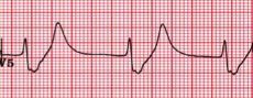
Picture
Management:
For patients with ESRD on hemodialysis, dialysis is the definitive treatment for hyperkalemia. 
Typical management of hyperkalemia (for K>6.0 or EKG changes) includes**:
• Calcium (either IV calcium gluconate or calcium chloride), which stabilizes cellular membranes by antagonizing the effect of potassium on membrane potential. Usually reserved for K>7.0 or EKG changes. 
• Insulin to promote the movement of potassium into cells.
• Albuterol and Sodium bicarbonate can also be used to promote the movement of potassium into cells depending on potassium levels.
• Kayexalate (Sodium polystyrene sulfonate) can be used to promote potassium elimination from the body, however this has no role in the acute management of hyperkalemia with EKG changes.
**Caution in patients with DKA because total body potassium may actually be low and aggressive management of hyperkalemia can result in hypokalemia**

By: Krystin Thomas, PGY1

Resources:
• Stone, CK. Fluid, Electrolyte, and Acid-Base Emergencies. In Current diagnosis & treatment emergency medicine. 6th ed. New York: McGraw-Hill; 2008. 
• Burns, E. Hyperkalemia [Web log post] Retrieved August 28, 2015, from http://lifeinthefastlane.com/ecg-library/basics/hyperkalaemia/

0 Comments

Your comment will be posted after it is approved.


Leave a Reply.

    EKG Challenge

    Picture

    Author

    ER residents on Dr. Littmann's cardiology service present an interesting EKG and core concepts from Dr. Littmann.

    Subscribe to Blog
    Littmann's EKG Pearls
    Videos Of Littmann's Lectures

    Archives

    January 2016
    November 2015
    October 2015
    September 2015
    August 2015
    July 2015
    June 2015
    May 2015
    April 2015

    Categories

    All
    Afib
    AIVR
    Aortic Stenosis
    Atrial Flutter
    AVR Sign
    Bifascicular Block
    Cardiac Tamponade
    CHF
    Dextrocardia
    HCOM
    High Lateral MI
    Hyperkalemia
    LAFB
    Paced Rhythms
    Pulmonary Hypertension
    PVC
    Respiratory Distress
    Short QT
    South African Flag Sign
    STEMI
    WPW

    Disclaimer: All EKG's and images are the sole property of CMC Emergency Medicine Residency and cannot be reproduced without written consent.  Patient identifiers have been redacted/changed or patient consent has been obtained.  Information contained in this blog is the opinion of the authors and application of material contained in this blog is at the discretion of the practitioner to verify for accuracy.

    RSS Feed

Disclaimer: All original material and images included on this website are the sole property of CMC EM Residency and cannot be used or reproduced without written permission.  Information contained on this website is the opinion of the authors and does not necessarily represent the official opinion of Atrium Health or Carolinas Emergency Medicine Residency. 


For Health Care Providers:  Every effort is made to provide the most up to date evidence based medicine.  However, this content may not necessarily reflect the standard of care and application of material contained on this website is at the discretion of the practitioner to verify for accuracy.


For the Public:  This content is not intended to be a substitute for professional medical advice, diagnosis, or treatment.  Relying on information contained on this website is done at your own risk.  Do not disregard professional medical advice or delay seeking care secondary to content on this website.  Call 911 or seek a medical professional immediately for any medical emergencies.
Like us on Facebook or
follow us on Twitter/Instagram

Contact Us:

Department of Emergency Medicine
Medical Education Building., Third floor
1000 Blythe Blvd.
Charlotte, NC 28203

Telephone: 704-355-3658 
Fax: 704-355-7047
​
  • Prospective Applicants
    • 2020 MATCH
    • About CMC
    • Our Curriculum
    • Our Residents
    • Our Fellowships
    • Program Leadership
    • Explore Charlotte
    • Official Site
  • ROTATING STUDENTS
    • Prospective Visiting Students
    • UNC Students
    • Healthcare Disparities Externship
    • Current Students
  • Current Residents
    • Airway Lecture!
    • PGY - 1
    • PGY - 2
    • PGY - 3
    • Simulation Reading
    • Blogs >
      • EM GuideWire
      • CMC ECG Masters
      • Core Concepts
      • #FOAMed
      • Cardiology Blog
      • Dr. Patel's Coding Blog
      • Global Health Blog
      • Ortho Blog
      • Pediatric Emergency Medicine
      • Tox Blog
    • Board Review
    • Journal Club
    • Resident Wellness
    • Resident Research
  • Top 20
  • Chiefs Corner
    • Schedules >
      • Conference/Flashpoint
      • Block Schedule
      • ED Shift Schedule
      • AEC Moonlighting
      • Journal Club/OBP/Audits Schedule
      • Simulation
    • Individualized Interactive Instruction
    • Evaluations/Interview Season
    • Contact Info
    • Resume Builder