CMC COMPENDIUM
  • RESIDENCY
    • About CMC
    • Curriculum
    • Benefits
    • Explore Charlotte
    • Official Site
  • FELLOWSHIP
    • EMS
    • Global EM
    • Pediatric EM
    • Toxicology >
      • Tox Faculty
      • Tox Application
    • (All Others)
  • PEOPLE
    • Program Leadership
    • PGY-3
    • PGY-2
    • PGY-1
    • Alumni
  • STUDENTS/APPLICANTS
    • Medical Students at CMC
    • EM Acting Internship
    • Healthcare Disparities Externship
    • Resident Mentorship
  • #FOAMed
    • EM GuideWire
    • CMC Imaging Mastery
    • Pediatric EM Morsels
    • Blogs, etc. >
      • CMC ECG Masters
      • Core Concepts
      • Cardiology Blog
      • Dr. Patel's Coding Blog
      • Global Health Blog
      • Ortho Blog
      • Pediatric Emergency Medicine
      • Tox Blog
  • Chiefs Corner
    • Top 20
    • Current Chiefs
    • Schedules >
      • Conference/Flashpoint
      • Block Schedule
      • ED Shift Schedule
      • AEC Moonlighting
      • Journal Club/OBP/Audits Schedule
      • Simulation
    • Resources >
      • Fox Reference Library
      • FlashPoint
      • Airway Lecture
      • Student Resources
      • PGY - 1
      • PGY - 2
      • PGY - 3
      • Simulation Reading
      • Resident Wellness
      • Resident Research
      • Resume Builder
    • Individualized Interactive Instruction

Something Seems Backwards Here...

5/1/2015

0 Comments

 
HPI:  Approximately 30 year old obese gentleman with 15 pack year smoking history presents with 2 days SOB, cough productive of yellow sputum, and occasional chest pain. He has a family history significant for MI at a young age so an EKG is obtained.

EKG:
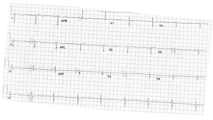
Picture
EKG Interpretation:
Heart rate is <60 and regular. This EKG is not in sinus rhythm. P waves are inverted in I and upright in avR.  There is significant axis deviation with QRS down going in lead I and II.  QRS normal width.  Abnormal R wave progression with QRS upgoing in lead V1 and R wave highest in V1.  No ST changes.  QT interval within normal.  Inverted T waves in V2-V5.

Discussion:
Differential for inverted P waves in lead 1 should always include the following:
  • Limb lead reversal
  • Ectopic atrial rhythm
  • Atrial paced rhythm
  • Dextrocardia

In dextrocardia the QRS is down going in lead I and the R wave progression is the opposite. R wave is highest in V1. The net vector is no longer going towards the left leg.  This patient has dextrocardia

Management:
How do you confirm this?
1. Reverse all the leads, chest and limb leads and repeat the EKG. This will allow you to properly interpret any abnormalities on the EKG.  
2. Obtain a chest x-ray to confirm dextrocardia
3. Inform patient of the diagnosis and if possible give patient a copy of his EKG or make a note in the patient chart.
4. If defibrillating a patient with dextrocardia, remember to place the pads in reverse position.

Dextrocardia Pearls:
There are two main types of dextrocardia:  dextrocardia of embryonic arrest and dextrocardia situs inversus.  In dextrocardia embryonic arrest, the heart is simply found more right in the chest cavity than normal.  In dextrocardia situs inversus, the heart is a mirror image situated on the right.  If all visceral organs are inverted, it is called dextrocardia situs inversus totalis.   Dextrocardia has a prevalence of approximately 1 in 12,000 people.
0 Comments

    EKG Challenge

    Picture

    Author

    ER residents on Dr. Littmann's cardiology service present an interesting EKG and core concepts from Dr. Littmann.

    Subscribe to Blog
    Littmann's EKG Pearls
    Videos Of Littmann's Lectures

    Archives

    January 2016
    November 2015
    October 2015
    September 2015
    August 2015
    July 2015
    June 2015
    May 2015
    April 2015

    Categories

    All
    Afib
    AIVR
    Aortic Stenosis
    Atrial Flutter
    AVR Sign
    Bifascicular Block
    Cardiac Tamponade
    CHF
    Dextrocardia
    HCOM
    High Lateral MI
    Hyperkalemia
    LAFB
    Paced Rhythms
    Pulmonary Hypertension
    PVC
    Respiratory Distress
    Short QT
    South African Flag Sign
    STEMI
    WPW

    Disclaimer: All EKG's and images are the sole property of CMC Emergency Medicine Residency and cannot be reproduced without written consent.  Patient identifiers have been redacted/changed or patient consent has been obtained.  Information contained in this blog is the opinion of the authors and application of material contained in this blog is at the discretion of the practitioner to verify for accuracy.

    RSS Feed

Powered by Create your own unique website with customizable templates.
  • RESIDENCY
    • About CMC
    • Curriculum
    • Benefits
    • Explore Charlotte
    • Official Site
  • FELLOWSHIP
    • EMS
    • Global EM
    • Pediatric EM
    • Toxicology >
      • Tox Faculty
      • Tox Application
    • (All Others)
  • PEOPLE
    • Program Leadership
    • PGY-3
    • PGY-2
    • PGY-1
    • Alumni
  • STUDENTS/APPLICANTS
    • Medical Students at CMC
    • EM Acting Internship
    • Healthcare Disparities Externship
    • Resident Mentorship
  • #FOAMed
    • EM GuideWire
    • CMC Imaging Mastery
    • Pediatric EM Morsels
    • Blogs, etc. >
      • CMC ECG Masters
      • Core Concepts
      • Cardiology Blog
      • Dr. Patel's Coding Blog
      • Global Health Blog
      • Ortho Blog
      • Pediatric Emergency Medicine
      • Tox Blog
  • Chiefs Corner
    • Top 20
    • Current Chiefs
    • Schedules >
      • Conference/Flashpoint
      • Block Schedule
      • ED Shift Schedule
      • AEC Moonlighting
      • Journal Club/OBP/Audits Schedule
      • Simulation
    • Resources >
      • Fox Reference Library
      • FlashPoint
      • Airway Lecture
      • Student Resources
      • PGY - 1
      • PGY - 2
      • PGY - 3
      • Simulation Reading
      • Resident Wellness
      • Resident Research
      • Resume Builder
    • Individualized Interactive Instruction