|
HPI: Teenage female with SOB, presyncope, and palpitations x 1 hour. HR is 220 in triage. BP 118/84, RR 28, SpO2 96%. Alert and oriented. EKG: 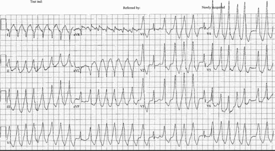 Question: What arrhythmia is this? How you would treat this rhythm in a stable patient. What if the patient were unstable? ECG Interpretation: Rate of 173/min. Irregularly irregular rhythm, wide complex tachycardia with intermittent narrow complexes and changing of QRS morphologies. No LBBB or RBBB pattern. Diagnosis: Afib with WPW Let’s discuss the interpretation of irregularly irregular Wide Complex Tachycardias (WCT), and then we will discuss the treatment. At first glance, it is difficult to assess the regularity, but with some scrutiny, you can see that the rhythm is irregular. The differential diagnosis of irregularly irregular WCT is essentially limited to three main dysrhythmias. Dr. Littmann calls these FBI: Fast, Broad, and Irregular:
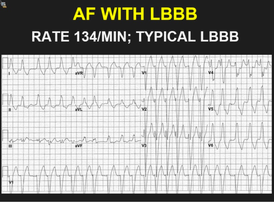 The above ECG is an irregularly irregular WCT meeting the criteria for LBBB as below (upgoing in I, deep S waves in V1). Note the rate of 134. Fast, but not extremely fast, indicating likely transmission through the AV node. 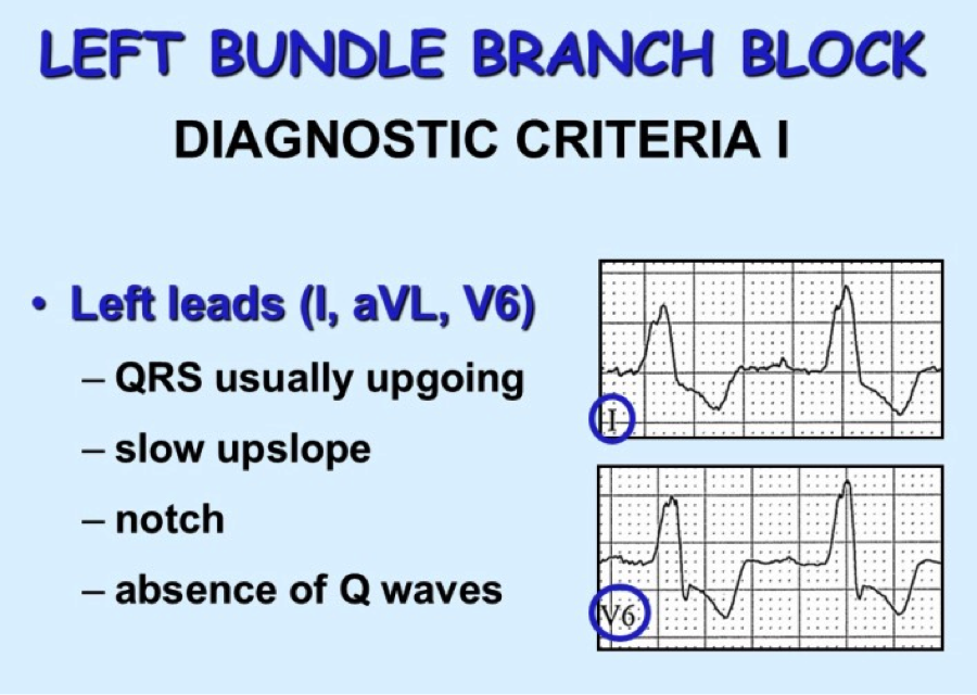 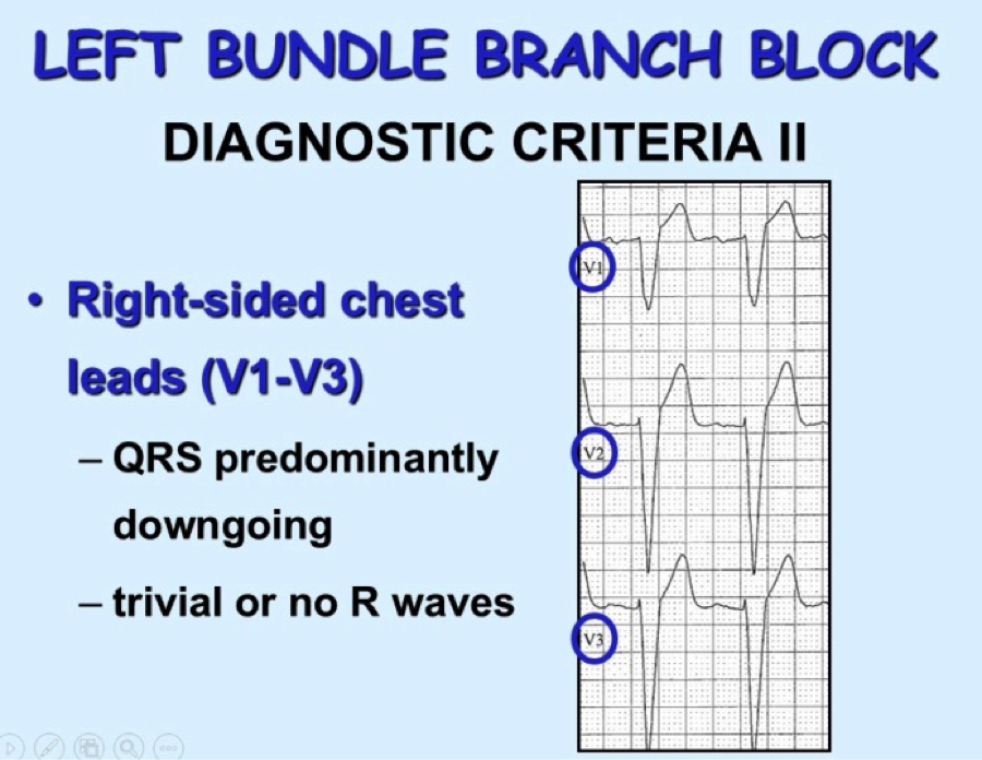 Consider RBBB, looking for triphasic QRS, with rSR’ and 2nd upgoing phase wider and taller than first upgoing phase as shown in these images: 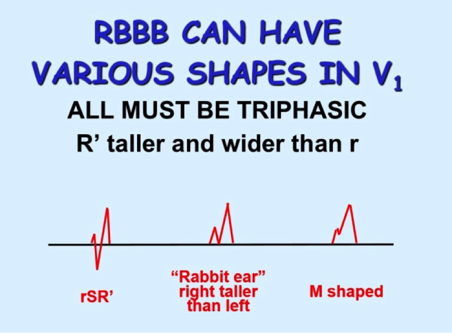 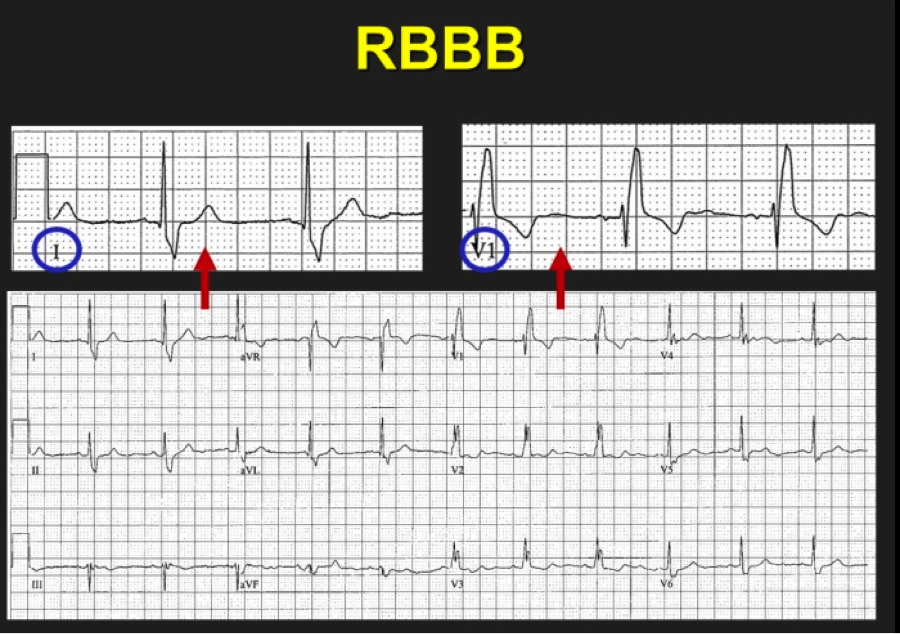 So we have scrutinized our irregularly irregular WCT and found no evidence of BBB pattern. We are now left with Afib with WPW. Why is this important? The treatment is completely different from regular WCT (usually Vtach) and Afib with BBB pattern. Treatment of FBI, Fast Broad and Irregular:
Key Points:
As a reminder for recognizing WPW in the non-tachycardic patient’s ECG: 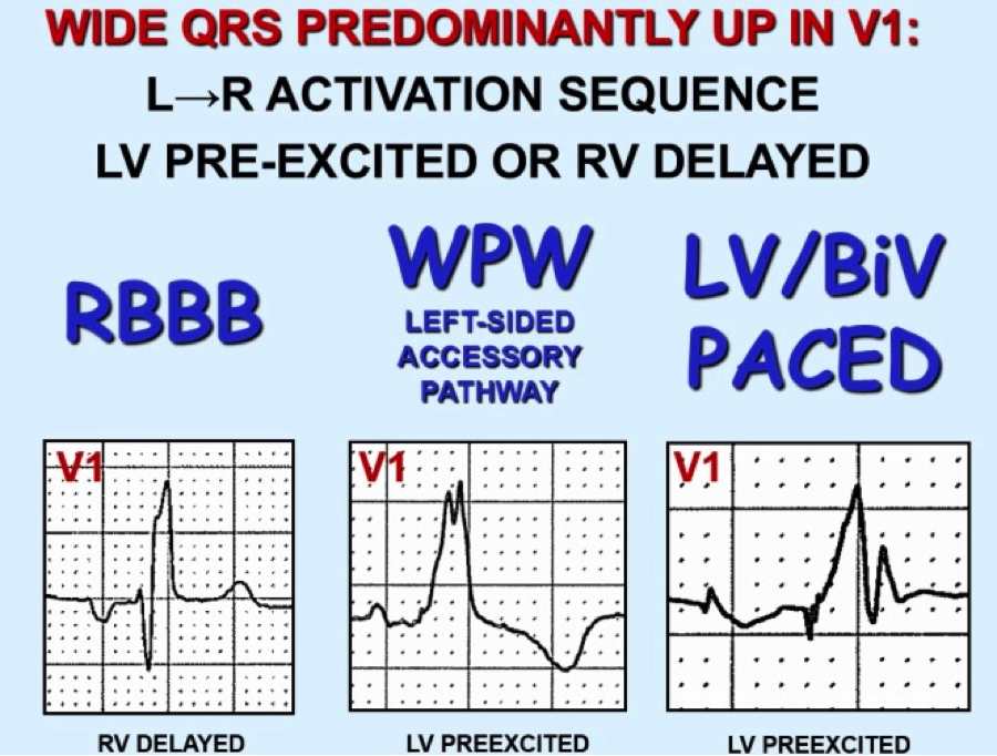 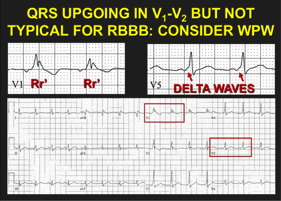 References:
By: Dean Tanner, PGY1 EM
0 Comments
HPI: Patient is a 54-year-old male presenting with palpitations and near syncope. Patient states that he was told he had a “heart problem” as a child. He was diagnosed with atrial fibrillation some 10+ years ago. EKG: 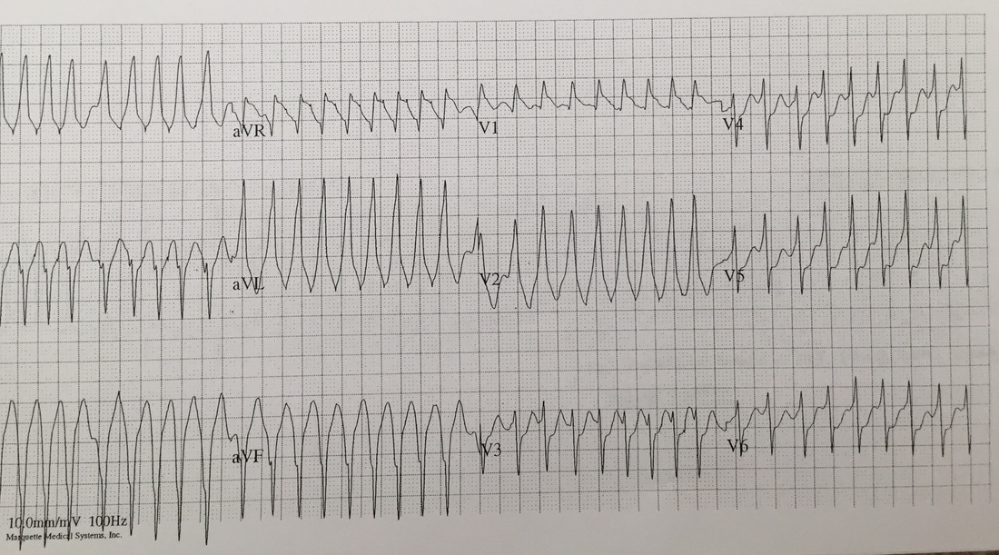 EKG Interperitation:
Rate estimated to be in the high 100s and irregularly irregular. QRS is wide. There are no appreciable P waves. Does not fit any typical bundle branch pattern. Discussion: Is this V tach? No. The patient has a wide complex irregulary irregular waveform. This is MOST likely to be a patient with WPW and a-fib. This is easily confused with ventricular tachycardia. How to differentiate: · Irregularly irregular · Rapid · Wide complex · Does not fit bundle branch pattern · No P waves Treatment: · DO NOT give: adenosine, verapamil, diltiazem, digoxin, beta blocker or amio o Due to the presence of accessory pathway blocking down the AV node, may cause the accessory pathway to become the primary driver of conduction. · Treated with IV procainamide if clinically stable. Dose is 15-16 mg/kg given at a rate no faster than 50 mg/min. · If unstable: Immediate DC cardoversion · Patients will require admission with likely EP study. By Dr Andrew Puchiaty |
EKG Challenge
AuthorER residents on Dr. Littmann's cardiology service present an interesting EKG and core concepts from Dr. Littmann. Archives
January 2016
Categories
All
Disclaimer: All EKG's and images are the sole property of CMC Emergency Medicine Residency and cannot be reproduced without written consent. Patient identifiers have been redacted/changed or patient consent has been obtained. Information contained in this blog is the opinion of the authors and application of material contained in this blog is at the discretion of the practitioner to verify for accuracy.
|
 RSS Feed
