|
Case Presentation: You are working in a Palestinian refugee clinic in Lebanon where a 3-year-old male presents to your clinic for high fevers for 1 day. The patient’s mother also states that the child has had a cough, runny nose, and red eyes. The patient and his family recently relocated to Lebanon from Aleppo, Syria. ROS: Otherwise negative PMH: Previously healthy PSH: Negative FH: Non-contributory Immunization History: Unable to seek regular primary care for the past 2 years secondary to conflict in Syria Allergies: None Physical Exam: Vital Signs: Temp = 39 C, HR = 150, BP = 90/50, RR = 34, O2 Sat = 96% RA General: Warm to the touch, appears tired, lethargic, cries during exam, but comforted by mother Skin: Cheeks are erythematous, no rash appreciated HEENT: Bilateral conjunctivitis, dry oral mucosa appreciated with blue-gray macules with erythematous base on buccal and gingival mucosa Neck: Spotty cervical lymphadenopathy, no meningeal signs Lungs: Course breath sounds bilaterally with upper airway noises CV: Tachycardia Abd: Soft, non-tender, non-distended Ext: No acute findings Neuro: Appears sleepy, but arousable and consolable by mother Differential Diagnosis
Work-Up
Diagnosis = MEASLES Measles
Etiology
Pathophysiology
History
Physical Exam
Complications
Treatment
Vaccinations
For more information:
http://www.who.int/immunization/diseases/measles/en/ http://refbooks.msf.org/msf_docs/en/measles/measles_en.pdf
0 Comments
Case Presentation A 13 yo boy presents to the clinic that you are working at in Cameroon. According to his mother, he started having watery diarrhea earlier in the day and has proceeded to have at least ten large bowel movements since that time. He has had two episodes of non-bloody, non-bilious diarrhea and has had tactile fever. He has been too weak to take in much liquid but has had a few intermittent sips of water. He has become progressively more fatigued throughout the day and was brought into the clinic by his mother and a few other family members. He has had no cough, congestion, or complaints of headache. A few other people in his community have had similar symptoms over the past week including his aunt and uncle. Of note, he has only urinated once since yesterday evening. Vital signs Temp: 99.4, HR: 138, RR: 15, BP: 90/60, Sats: 97% on room air Photo Credit: http://cameroon-info.net/stories/0,29767,@,cholera-epidemic-persists-in-cameroon.html Physical exam General: arouses to stimulation but sleepy in appearance, can answer questions HEENT: Normocephalic, sunken eyes bilaterally, TMs clear, nares clear without discharge, pharynx without erythema Neck: no stiffness, full ROM LAD: no cervical or axillary LAD Respiratory: clear to auscultation bilaterally, no crackles, no retractions, no tachypnea Cardiovascular: tachycardic, no murmur, peripheral pulses are rapid, no edema noted GI: abdomen is non-distended, soft, mildly tender to palpation, hyperactive bowel sounds Neuro: unable to walk without support, decreased strength in upper and lower extremities bilaterally Differential Diagnosis Escherichia coli Vibrio cholerae Campylobacter Norovirus Non-typhoidal Salmonella enterica Labs CMP Stool studies Discussion Cholera is an intestinal infection that leads to secretory diarrhea and is caused by the gram-negative bacteria V. cholera. It has more than 200 serotypes and produces an enterotoxin that leads to large volumes of fluid loss from the duodenum and jejunum. The colon is unable to reabsorb this large volume of fluid and can cause prolific amounts of diarrhea. People primarily become infected with cholera through ingestion of contaminated food and water, by ingesting undercooked shellfish/oysters, and by living in conditions with poor sanitation, which are more likely to have contaminated water supplies. Many adults in a community where cholera is endemic can be asymptomatic carriers and shed the bacteria in the their stools for two to three weeks. The incubation period is typically 24-48 hours but can be up to five days. Cholera is typically diagnosed clinically although stool studies and electrolyte panels can be sent to determine diagnosis and degree of dehydration and acidosis. Physical exam findings consistent with dehydration include sunken eyes, tachycardia, hypotension, poor skin turgor, delayed capillary refill, lethargy, and weak pulses. Management is focused on rehydration and correction of electrolyte abnormalities with fluid resuscitation. If rehydration is not performed in an adequate time frame, the disease can often be fatal. Examples of cholera cots, for management of large volume diarrhea
 Case Presentation In Port au Prince, Haiti a 15 year old male presents to the hospital with 10 days of fever, abdominal pain and decreased appetite. He had been seen by a local physician earlier in the week, was reassured and sent home. He now returns to a local hospital with increased fever, worsening abdominal pain and mental status changes. Physical Exam: Temp 38.9 HR: 85 RR: 20 BP: 110/70 02 sats: 95% on room air General: Toxic appearing male, difficult to arouse Heent: NCAT, mucous membranes dry, no neck stiffness Lungs: CTA bilaterally CV: RRR, no m/r/g Abd: Diminished bowel sounds, firm, distended, diffusely tender to palpation Skin: no rash Differential Diagnosis: Bacterial Gastroenteritis Appendicitis Malaria Typhoid Fever Amebiasis (Entamoeba histolytica with amebic colitis) Dengue Fever Leishmaniasis Initial Evaluation: CBC with diff CMP KUB Blood culture if available Rapid diagnostic malaria test Diagnosis: Typhoid Fever (Enteric Fever) Case continuation: The patient had evidence of intestinal perforation and went emergently to the OR for resection and repair. He was started on IV ceftriaxone and Flagyl. His recovery was slow post-operatively due to the delay in seeking care from the time he perforated. Typhoid Fever Typhoid Fever: There are over 27 million cases worldwide with 200,000 associated deaths annually. In endemic areas, it is more common in children and young adults. Typhoid fever is caused by Salmonella typhi and Salmonella paratyphi A, B and C which is transmitted by the ingestion of contaminated food and water. The most important reservoirs of infection are the convalescent carriers (Patients with acute disease continue to carry the bacteria for weeks to months following treatment before spontaneous resolution) and chronic carriers (asymptomatic patients who continue to carry the bacteria in the gall bladder or urine and intermittently shed the bacteria). Clinical Manifestations: Typhoid is a progressively febrile illness usually 1-3 weeks after ingestion of the bacteria. Non-specific abdominal pain, headache, nonproductive cough and diarrhea or constipation are common as are constitutional symptoms (fever, chills, anorexia, malaise). A relative bradycardia may be seen. In severe cases, patients may present with septic shock, altered mental status, DIC and lung disease. Children have pneumonia, febrile seizures and neuropsychiatric symptoms more often than adults. Clinical Progression:
Pathogenesis: S. typhi penetrate the submucosa of the small bowel where bacteria proliferate leading to inflammation and hypertrophy of peyers patches. Later in disease, this hypertrophy and resultant necrosis of submucosal tissues is probably responsible for the abdominal pain and subsequent bowel perforation. Bacteria enter the bloodstream via the thoracic duct and disseminate to many parts of the body including the bone marrow and gall bladder. Eventually bacteria is rereleased into the bloodstream, this second bacteremia corresponds to the onset of symptoms. Diagnosis: Isolation of the organism in the setting of clinical illness: Blood culture (positive in 40-80%), Aspirates from abscesses, CSF, Bone marrow for culture and Stool culture (positive in 30-40%) Treatment: Adults: Ciprofloxacin x 7-10 days or Ceftriaxone/Cefixime x 10-14 days
Outcomes/Complications: Successful treatment results in clinical improvement within 3-5 days Complications:
Vaccines: Live attenuated oral vaccine: Ty21a 3 doses over 5 days with a booster every 5 years (>6yo) Purified Vi Antigen vaccine IM: Booster every 3 years (>2yo) For More Information: http://www.who.int/rpc/TFGuideWHO.pdf http://www.cdc.gov/nczved/divisions/dfbmd/diseases/typhoid_fever/technical.html Case Presentation: 3 year old female presents to a district hospital in Dar es Salaam for concern over breathing fast and decreased energy. Approximately 2 weeks ago the child was pushed into a hole and then development progressive dyspnea. Child continued to worsen with no intervention and treatments over a 2 week period. The patient has no significant past medical or surgical history and no known allergies. Physical Exam: Vital Signs: HR 150 BP 92/60 RR 40 Temp 37.6 C PO O2 Sats 97-100% Awake and alert, noted to be leaning to the left Tachypneic Swelling over L chest ant L scapula Lung sounds absent on L side of chest Trachea is deviated to the right Apical impulse is shift to the right Differential Diagnosis: Hemothorax Pnemothorax Pneumonia Empyema Pleural Effusion Bronchial obstruction/atelectasis Tests: Chest XR IVF What if you had no immediate access to radiological imaging? Remember the use of bedside ultrasound! 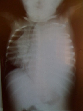 Concern for Empyema Next Steps: Chest tube placement Antibiotics Sedation Local Anesthesia Sterile technique 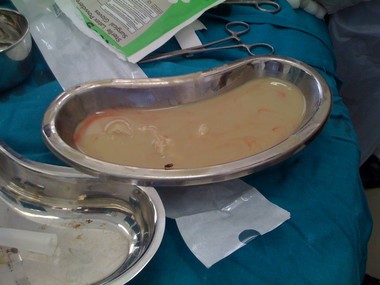 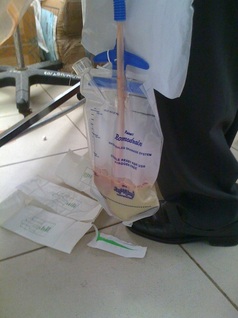 Reassessment:
Child’s tachypnea has improved after chest tube placement and nearly 1 liter of pus was drained from the chest. Case Pearls: Utilization of bedside ultrasound Remember your ultrasound skills and abilities when you are working in a setting with limited resources. If you cannot obtain immediate chest x-ray, you can use your bedside ultrasound to evaluate the chest to diagnosis pneumothorax, hemothroax, empyema, pneumonia, and much more. See tutorial on thoracic ultrasound or review article on emergency ultrasound of the chest. Empyema Empyema is inflammatory fluid and debris within the intrapleural space, usually results from an untreated bacterial pneumonia. However, an empyema can also be caused by thoracic trauma (like our case). Other causes include extension of mediastinal or abdominal infection or iatrogenic causes (chest tube placement or surgery). Chest tube placement Before performing any procedure always consider what type of analgesia and sedation the patient will need. Consider the patient’s vital signs/stability and the urgency of procedure when selecting sedation plan. To review chest tube placement watch the following videos. Thanks Dr. Mike Runyon for the case submission. History of Present Illness: A 24-month-old boy presents to the clinic you are working in rural Tanzania. He weighs 8.5kg. He was brought to clinic today by his mother because he has had decreased energy for a few weeks and has been very irritable and fussy. She has reports that he has had a “swollen belly for a long time”. He typically eats a chapatti for breakfast and a small bowel of local porridge for lunch and dinner. His other notable symptoms include multiple episodes of diarrhea for one week that is non-bloody and watery and a fever and cough that he has had for a few days. In addition, his oral intake has decreased the past few days and he was becoming sleepier at home today. Physical Exam: Vital Signs: Temp: 38.7 HR: 150 RR: 35 BP: 90/70 Pulse ox: 95% on room air General: Patient is tired in appearance but arousable on examination. HEENT: Normocephalic, TMs clear, Hair is reddish in appearance and ruddy colored. He has bilateral periorbital edema. His face is swollen. Neck: Generalized LAD. Lungs: CTAB, no wheezing, no retractions noted Heart: tachycardia, no murmur, peripheral pulses are 2+ Abdomen: firm, distended, nontender, has bowel sounds, liver is enlarged and down 4 cm. Skin: Areas of dry peeling skin on extremities bilaterally with patches of hyperpigmentation. Extremities: generalized edema with 3+ edema in arms and legs bilaterally. Pedal edema is pitting. 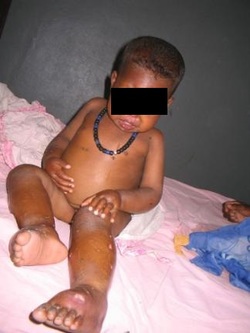 Questions: What is the most likely diagnosis of this patient? What physical exam findings support the diagnosis? What is the typical treatment for the patient’s condition? Does this patient meet inpatient treatment criteria? Answers:
What is the most likely diagnosis of this patient? This patient has a condition called kwashiorkor. Kwashiorkor is a form of malnutrition that is secondary to inadequate protein and micronutrient deficiency. The inadequate amount of protein in the diet not only leads to malnutrition but also results in hypoalbuminemia that leads to third spacing and accumulation of extravascular fluid causing the edematous presentation in patients. This leads to physical exam findings such as moon facies and pitting edema. Typically the patient also has concurrent zinc deficiency that can cause dermatitis leading to desquamation of the skin and either hyper or hypopigmentation. Hepatomegaly is another common physical exam finding that is caused from fatty infiltration of the liver. These patients are often apathetic, listless, and irritable. Hair often becomes dry and brittle and sometimes changes to a ruddy, orange color. What physical exam findings support the diagnosis? Typically Kwashiorkor is a clinical diagnosis based on physical exam findings, weight for age, and history. Other diagnosis for generalized edema should be considered, but in a rural area known to have a large population with malnutrition, this is typically the most likely diagnosis. Patients typically have electrolyte abnormalities, vitamin deficiencies, and often will be anemic. If the clinic/hospital you are working in has the capability to send for labs, a CMP, CBC, Mg, and Phos would be useful. Often these children present with dehydration with concurrent hypovolemic shock. Children with kwashiorkor are prone to developing infection and can also present with fever due to septic shock, pneumonia, cellulitis due to infection of sloughing skin, or other infections. Alternatively, these children can also be hypothermic due to infection, hypoglycemia, or lack of fat/muscle mass. What is the typical treatment for the patient’s condition? Treatment is many fold and is directed primarily at correcting electrolyte/glucose abnormalities, rehydrating if dehydrated, treating with antibiotics if infection is suspected, deworming, correcting vitamin/micronutrient deficiencies, and providing adequate nutrition to promote weight gain. Patients should be monitored closely during stabilization process. Most hospitals have a protocol for malnutrition that assists in helping children to gain weight slowly and safely. Does this patient meet inpatient treatment criteria? Children meet criteria for inpatient treatment if they have a poor appetite and fail an appetite test in clinic, if they have severe medical complications, if there is edema present on physical exam, or if they are failing outpatient management. Medical complications include intractable vomiting, signs concerning for severe dehydration, fever or hypothermia, signs of severe lower respiratory tract infection such as tachypnea/hypoxia/respiratory distress, severe anemia, or altered mental status/lethargy. |
Global health
Blog posts from the resident and faculty physicians of the Carolinas Medical Center global health interest group.
Archives
April 2016
Categories |
 RSS Feed
