|
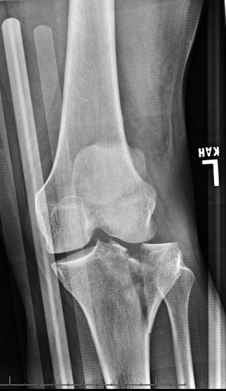
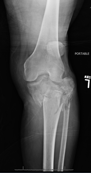
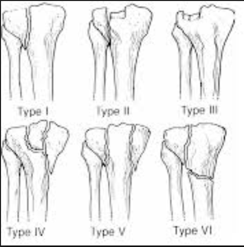
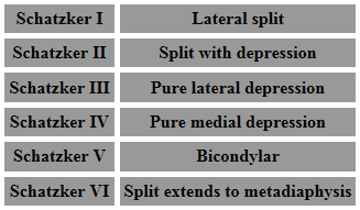
HPI: Patient 1: Restrained MVC. Presented with knee pain and swelling. Patient 2: Fall from height and chief complaint of shoulder and knee pain. Physical Exam: Patient 1: Swollen left knee with tenderness to palpation. Patient 2: Knee visibly deformed, swollen, and bruised. Small open deformity w/ active bleeding. High risk for compartment syndrome. Imaging: Patient 1: Schatzker type 2 fracture (see table below for classifications of Schatzker fracture) Patient 2: Schatzker type 6 fracture
Dispo: Patient 1: Admitted. Splinted w/ ORIF as inpatient. Patient 2: Admitted. Splinted w/ closed reduction and external fixation. Plan for definitive repair in ~2 weeks after soft tissue swelling has subsided. Treatment: 1) Hinged knee brace w/ passive ROM - for patients w/ minimally displaced or split depressed fractures or minimal baseline mobility. 2) External fixation - for patient w/ open or comminuted fractures or significant soft tissue swelling. 3) ORIF - for >3mm articular step off or varus/valgus instability. Types of Schatzker Fractures:
0 Comments
Leave a Reply. |
Orthopedics Blog
AuthorCMC ER Residents Archives
June 2018
Categories
All
Disclaimer: All images and x-rays included on this blog are the sole property of CMC EM Residency and cannot be used or reproduced without written permission. Patient identifiers have been redacted/changed or patient consent has been obtained. Information contained in this blog is the opinion of the author and application of material contained in this blog is at the discretion of the practitioner to verify for accuracy.
|
 RSS Feed
